Mouse Nogo Receptor/NgR Antibody Summary
Cys27-Ser447
Accession # Q99PI8
Applications
Please Note: Optimal dilutions should be determined by each laboratory for each application. General Protocols are available in the Technical Information section on our website.
Scientific Data
 View Larger
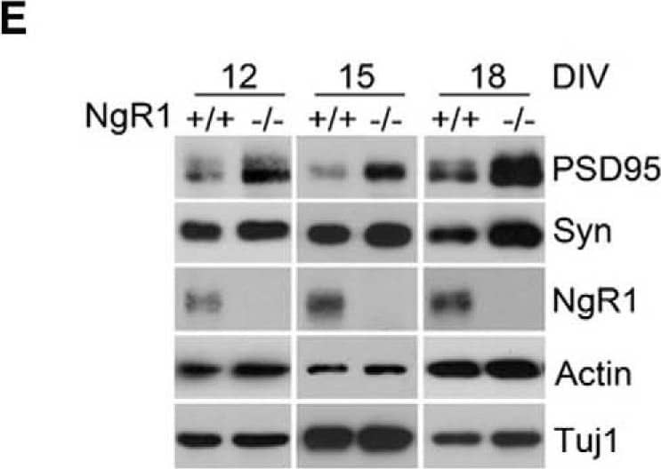
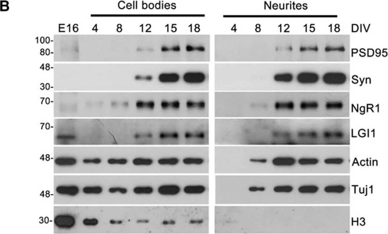
Detection of Mouse Nogo Receptor/NgR by Western Blot NgR1 and LGI1 regulate synaptic proteins in cortical neurons in vitro.A, Twiss filter schematic showing culture system to coculture hippocampal neurons with astrocytes and separate neuronal processes from cell bodies. Hippocampal neurons seeded on filters with a pore size 1 µm that cell bodies will not pass through. Axons and dendrites grow on the filter tops and extend down onto the filter bottom. Astrocytes are seeded on the bottom of the well to provide growth factors. B, Time course of lysates from hippocampal neurons grown on filters suspended over an astrocyte feeder layer for the times indicated. The first lane in the left panel labeled E16 is a sample of hippocampal neurons lysed directly after dissociated before plating. Lysates from filter tops including cell bodies and processes are on the left. Lysates of the filter bottoms containing axons and dendrites but no cell bodies are on the right. Antibodies used to probe the lysates are indicated on the right. Histone-3 (H3), a structural protein found in chromatin and present only in the nucleus is detected only in the cell body lysates. C, Lysates from filter bottoms containing axons and dendrite but not cell bodies from LGI1+/+ and LGI1-/- littermates of cortical cultures grown for the indicated number of DIV. D, Quantification of PSD95 levels relative to actin levels and normalized to WT controls in LGI1 samples at 12, 15, and 18 DIV, n = 3 separate experiments. E, Western blottings of lysates from filter bottoms of NgR1+/+ and NgR1-/- cortical cultures harvested at 12, 15, or 18 DIV synaptic markers, Syn and PSD95. Actin and Tuj1 are loading controls. F, Quantification of PSD95 relative to actin levels and normalized to WT controls in NgR1, n = 4 separate experiments. Significant differences are indicated on the graphs analysis was performed by two-way ANOVA with Bonferroni post hoc tests, **p < 0.01, *p < 0.05. Image collected and cropped by CiteAb from the following open publication (https://pubmed.ncbi.nlm.nih.gov/30225353), licensed under a CC-BY license. Not internally tested by R&D Systems.
Reconstitution Calculator
Preparation and Storage
- 12 months from date of receipt, -20 to -70 °C as supplied.
- 1 month, 2 to 8 °C under sterile conditions after reconstitution.
- 6 months, -20 to -70 °C under sterile conditions after reconstitution.
Background: Nogo Receptor/NgR
Nogo Receptor (NgR), also named reticulon 4 receptor, is a glycosylphosphoinositol (GPI)-anchored protein that belongs to the family of leucine-rich repeat (LRR) proteins (1). It is expressed predominantly in the central nervous systems in neurons and their axons. NgR plays an essential role in mediating axon growth inhibition induced by the structurally distinct myelin-derived proteins Nogo, myelin-associated glycoprotein (MAG), and myelin oligodendrocyte glycoprotein (Omgp) (2, 3). Human NgR cDNA encodes a 473 amino acid (aa) residue precursor with a 26 aa putative signal peptide, an LRR-type N-terminal region, eight LRR repeats, a cysteine-rich LRR-type C-terminal region, a GPI linkage domain and a 26 aa C-terminal propeptide that is removed in the mature form (1). All of the LRR domains within NgR are required for ligand binding and receptor oligomerization (4). NgR mediates its inhibitory actions by interacting with the p75 neurotrophin receptor (p75NTR), a tumor necrosis factor receptor superfamily (TNFRSF) member also known for modulating the activities of the Trk family of receptor tyrosine kinases, and for inducing apoptosis in neurons and oligodendrocytes (5). Upon ligand binding, NgR binds to and activates the p75NTR. The activated p75NTR then sequesters the Rho guanine dissociation inhibitor (Rho-GDI) away from Rho and allows Rho to change into the active GTP-bound state which can interact with signaling proteins to suppress axonal growth and regeneration (4). The truncated extracellular domain of NgR has been shown to bind the myelin-derived inhibitors and block inhibition of axon growth by myelin (6).
- Fournier, A.E. et al. (2001) Nature 409:341.
- GrandPre, T. et al. (2002) Nature 417:547.
- Wang, K.C. et al. (2002) Nature 420:74.
- Barton, W.A. et al. (2003) EMBO Journal 22:3291.
- Yamashita, T. and M. Tohyama (2003) Nature Neuroscience 6:461.
- Fournier, A.S. et al. (2002) J. Neurosci. 22:8876.
Product Datasheets
Citations for Mouse Nogo Receptor/NgR Antibody
R&D Systems personnel manually curate a database that contains references using R&D Systems products. The data collected includes not only links to publications in PubMed, but also provides information about sample types, species, and experimental conditions.
18
Citations: Showing 1 - 10
Filter your results:
Filter by:
-
Distinct Circuits for Recovery of Eye Dominance and Acuity in Murine Amblyopia
Authors: Stephany CE, Ma X, Dorton HM et al.
Curr Biol
-
Fast Regulation of GABAAR Diffusion Dynamics by Nogo-A Signaling
Authors: S Fricke, K Metzdorf, M Ohm, S Haak, M Heine, M Korte, M Zagrebelsk
Cell Rep, 2019-10-15;29(3):671-684.e6.
Species: Mouse
Sample Types: Whole Cells
Applications: Neutralization -
The Nogo Receptor Ligand LGI1 Regulates Synapse Number and Synaptic Activity in Hippocampal and Cortical Neurons
Authors: Rhalena A. Thomas, Julien Gibon, Carol X. Q. Chen, Sabrina Chierzi, Vincent G. Soubannier, Stephanie Baulac et al.
eNeuro
-
Nogo receptor 1 is expressed by nearly all retinal ganglion cellsPV expression in Figure 3
Authors: AM Solomon, T Westbrook, GD Field, AW McGee
PLoS ONE, 2018-05-16;13(5):e0196565.
Species: Mouse
Sample Types: Whole Tissue
Applications: IHC -
The nociceptin receptor inhibits axonal regeneration and recovery from spinal cord injury
Authors: Yuichi Sekine, Chad S. Siegel, Tomoko Sekine-Konno, William B. J. Cafferty, Stephen M. Strittmatter
Science Signaling
-
The soluble form of LOTUS inhibits Nogo receptor-mediated signaling by interfering with the interaction between Nogo receptor type 1 and p75 neurotrophin receptor
Authors: Y Kawakami, Y Kurihara, Y Saito, Y Fujita, T Yamashita, K Takei
J. Neurosci., 2018-02-09;0(0):.
Species: Mouse
Sample Types: Whole Cells
Applications: ICC -
Amyloid Beta Peptides Block New Synapse Assembly by Nogo Receptor-Mediated Inhibition of T-Type Calcium Channels
Authors: Yanjun Zhao, Sivaprakash Sivaji, Michael C. Chiang, Haadi Ali, Monica Zukowski, Sareen Ali et al.
Neuron
-
Regulation of axonal regeneration by the level of function of the endogenous Nogo receptor antagonist LOTUS
Authors: T Hirokawa, Y Zou, Y Kurihara, Z Jiang, Y Sakakibara, H Ito, K Funakoshi, N Kawahara, Y Goshima, SM Strittmatt, K Takei
Sci Rep, 2017-09-21;7(1):12119.
Species: Mouse
Sample Types: Tissue Homogenates
Applications: Western Blot -
LOTUS overexpression accelerates neuronal plasticity after focal brain ischemia in mice
Authors: H Takase, Y Kurihara, TA Yokoyama, N Kawahara, K Takei
PLoS ONE, 2017-09-07;12(9):e0184258.
Species: Mouse
Sample Types: Tissue Homogenates
Applications: Western Blot -
Erasure of fear memories is prevented by Nogo Receptor 1 in adulthood
Authors: Sarah M. Bhagat, Santino S. Butler, Jane R. Taylor, Bruce S. McEwen, Stephen M. Strittmatter
Molecular Psychiatry
-
Human NgR-Fc Decoy Protein via Lumbar Intrathecal Bolus Administration Enhances Recovery from Rat Spinal Cord Contusion
Authors: Xingxing Wang, Kazim Yigitkanli, Chang-Yeon Kim, Tomoko Sekine-Konno, Dana Wirak, Eric Frieden et al.
Journal of Neurotrauma
-
Cell type-specific Nogo-A gene ablation promotes axonal regeneration in the injured adult optic nerve.
Authors: Vajda F, Jordi N, Dalkara D, Joly S, Christ F, Tews B, Schwab M, Pernet V
Cell Death Differ, 2014-09-26;22(2):323-35.
Species: Mouse
Sample Types: Whole Tissue
Applications: IHC -
Plasticity of binocularity and visual acuity are differentially limited by nogo receptor.
Authors: Stephany C, Chan L, Parivash S, Dorton H, Piechowicz M, Qiu S, McGee A
J Neurosci, 2014-08-27;34(35):11631-40.
Species: Mouse
Sample Types: Whole Tissue
Applications: IHC -
LGI1 is a Nogo receptor 1 ligand that antagonizes myelin-based growth inhibition.
Authors: Thomas R, Favell K, Morante-Redolat J, Pool M, Kent C, Wright M, Daignault K, Ferraro GB, Montcalm S, Durocher Y, Fournier A, Perez-Tur J, Barker PA
J. Neurosci., 2010-05-12;30(19):6607-12.
Species: Human
Sample Types: Cell Lysates
Applications: Western Blot -
Soluble Nogo receptor down-regulates expression of neuronal Nogo-A to enhance axonal regeneration.
Authors: Peng X, Zhou Z, Hu J, Fink DJ, Mata M
J. Biol. Chem., 2009-11-09;285(4):2783-95.
Species: Rat
Sample Types: Cell Lysates
Applications: Western Blot -
Localization of an axon growth inhibitory molecule Nogo and its receptor in the spinal cord of mouse embryos.
Authors: Wang J, Wang L, Zhao H, Chan SO
Brain Res., 2009-10-13;1306(0):8-17.
Species: Mouse
Sample Types: Whole Tissue
Applications: IHC -
Reassessment of corticospinal tract regeneration in Nogo-deficient mice.
Authors: Lee JK, Chan AF, Luu SM, Zhu Y, Ho C, Tessier-Lavigne M, Zheng B
J. Neurosci., 2009-07-08;29(27):8649-54.
Species: Mouse
Sample Types: Tissue Homogenates
Applications: Western Blot -
Synaptic function for the Nogo-66 receptor NgR1: regulation of dendritic spine morphology and activity-dependent synaptic strength.
Authors: Lee H, Raiker SJ, Venkatesh K, Geary R, Robak LA, Zhang Y, Yeh HH, Shrager P, Giger RJ
J. Neurosci., 2008-03-12;28(11):2753-65.
Species: Rat
Sample Types: Cell Lysates
Applications: Western Blot
FAQs
No product specific FAQs exist for this product, however you may
View all Antibody FAQsReviews for Mouse Nogo Receptor/NgR Antibody
Average Rating: 4 (Based on 1 Review)
Have you used Mouse Nogo Receptor/NgR Antibody?
Submit a review and receive an Amazon gift card.
$25/€18/£15/$25CAN/¥75 Yuan/¥2500 Yen for a review with an image
$10/€7/£6/$10 CAD/¥70 Yuan/¥1110 Yen for a review without an image
Filter by:


