CMP-Cy3-Sialic Acid Summary
Key Benefits
Learn more about Fluorescent Glycan Labeling and Detection

|
Formula |
C52H73N10O25P1S3 |
|
Molecular Weight |
1413.25 Da |
|
Formulation |
Lyophilized with Tris, pH 8.0 |
|
Stability & Storage |
Store the unopened product at < -20 °C. Good for 12 months from date of receipt. |
Applications
- Fluorescent labeling with Cy3 of free glycans as well as glycoproteins and glycolipids.
- Fluorescent detection of specific glycan epitopes on the cell surface.
- Quantitation of the sialylation level of specific glycans.
- Together with CMP-Cy5-Sialic Acid, allows for dual labeling and detection of sialoglycans.
Key Features and Benefits:
- Excitation at 550 nm and emission at 570 nm, exhibits green fluorescent light under microscope.
- The fluorescent dye Cy3 is conjugated to the C9 position of the sialic acid.
- Can be directly introduced into glycoproteins and glycolipids via various sialyltransferases.
- Can be introduced to live cells for glycan imaging.
- Have minimum side-effect on target molecules.
- Very convenient and user-friendly.
For Details:
Wu et al., (2019) Glycobiology 29: 750-754.
Wu et al., (2020) Glycobiology 30:454-462.
Related Reagents
Click Chemistry
- Biotinylated-Alkyne (ES100)
- GDP-Azido-Fucose (ES101)
- CMP-Azido-Sialic Acid (ES102)
- UDP-Azido-GalNAc (ES103)
- UDP-Azido-GlcNAc (ES104)
- CMP-C9-Biotin-Sialic Acid (ES201)
- GDP-Cy5-Fucose (ES301)
- CMP-Cy5-Siaic Acid (ES302)
- GDP-Cy3-Fucose (ES401)
Enzymes and detection reagents
- Various sialyltransferases
- Various neuraminidases/sialidases
Data Examples
|
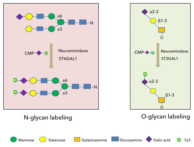
Strategies for N- and O- glycan labeling with CMP-Cy3-Sialic Acid. Only representative glycans are depicted here. Glycans can be labeled with or without neuraminidase treatment. A selected neuraminidase will remove existing sialic acids and create more labeling sites for a selected sialyltransferase. While Cy5 conjugated sialic acid can be recognized by most of known sialyltransferases, it is poorly recognized by any known neuraminidase, which allows for labeling in a single reaction. The pairing of a neuraminidase and a sialyltransferase is based on the compatibility between the two enzymes. For example, Recombinant C. perfringens Neuraminidase Protein (Catalog # 5080-NM) can remove both α2-3 and α2-6 linked sialic acids, and it can be paired with all α2-3 specific sialyltransferases (ST3GALs) and all α2-6 specific sialyltransfeases (ST6GALs). In the case that α2-8 linked sialic acid needs to be labeled, specific neuraminidase such as Recombinant M.viridifaciens Neuraminidase Protein (Catalog # 5084-NM) and α2-8 specific sialyltransferases (ST8SIAs) will be paired. |
|
Labeling bovine fetuin (Fet) using CMP-Cy3-Sialic Acid. Bovine fetuin was purified from crude fetuin by gel filtration. Samples of fetuin were labeled through O-glycans with Recombinant Human ST3GAL1 Protein (Catalog # 6905-GT) or N-glycans with Recombinant Human ST6GAL1 (aa 44-406) Protein (Catalog # 7620-GT). Control lanes contain all components but lack a labeling enzyme. Each lane contained 1 μg of fetuin except the controls. The control lanes contained 2 μg of protein. Samples exhibited greater labeling in the presence of Recombinant C. perfringens Neuraminidase Protein (Catalog # 5080-NM) (Neu, indicated with + and - signs), suggesting that both N- and O-glycans on fetuin were largely sialylated before labeling. Samples were separated on 4-20% gradient SDS-PAGE and imaged with TCE image (top panel) and a fluorescent imager (bottom panel). The bands of fetuin corresponding to the neuraminidase treated samples appear to be darker than those of without neuraminidase treatment because of the incorporation of Cy3. ST6GAL1 also showed some self-labeling in the presence of neuraminidase (indicted with an arrow in the bottom panel). M, Western blot molecular marker. |
|
Labeling recombinant MUC16 using CMP-Cy3-Sialic Acid. Samples were labeled through O-glycans with Recombinant Human ST3GAL1 Protein (Catalog # 6905-GT) or N-glycans with Recombinant Human ST6GAL1 (aa 44-406) Protein (Catalog # 7620-GT). Control lanes contain all components but lack a labeling enzyme. Each lane contained 1 μg of fetuin except the controls. The control lanes contained 2 μg of protein. Samples exhibited greater labeling in the presence of Recombinant C. perfringens Neuraminidase Protein (Catalog # 5080-NM) (Neu, indicated with + and - signs), suggesting that both N- and O-glycans on MUC16 were largely sialylated before labeling. In fact, by densitometry analysis on the fluorescent bands of MUC16 before and after neuraminidase treatment, the sialyation of the protein was measured to be 91.3% for O-glycans and 79.9 % for N-glycans. Samples were separated on 4-20% gradient SDS-PAGE and imaged with TCE (top panel) and a fluorescent imager (bottom panel). The bands of MUC16 corresponding to the neuraminidase treated samples appear to be darker than those of without neuraminidase treatment because of the incorporation of Cy3. M, Western blot molecular marker. |
Table 1. Guideline for using CMP-Cy5-Sialic Acid for sialoglycan labeling and detection
| Sialyltransferase | Catalog # | Substrate | CMP-Cy5-Sialic Acid Compatibility | Neuraminidase |
| ST3GAL1 | 6905-GT | O-glycan | yes | rCpNeuraminidase |
| ST3GAL2 | 7275-GT | O-glycan | yes | (Catalog # 5080-NM) |
| ST3GAL3 (coming soon) | 10554-GT | N- and O-glycan | yes | |
| ST3GAL4 (coming soon) | 10496-GT | N-glycan | yes | |
| ST3GAL5 | 8454-GT | Glycolipid | Not tested | |
| ST3GAL6 (coming soon) | 10591-GT | N-glycan | yes | |
| ST6GAL1 | 7620-GT | N-glycan | yes | |
| ST6GALNAC1 | 9154-GT | O-Glycan | yes | |
| ST6GALNAC2 | 6468-GT | O-glycan | yes | |
| ST6GALNAC4 | 6876-GT | O-glycan | Not tested | |
| ST6GALNAC5 | 6715-GT | Ganglioside | Not tested | |
| ST6GALNAC6 | 7425-GT | Ganglioside | Not tested | |
| ST8SIA1 | 6716-GT | Polysialic Acid | yes | rMvNeuraminidase |
| ST8SIA2 | 6590-GT | yes | (Catalog # 5084-NM) | |
| ST8SIA4 | 7027-GT | yes | ||
| ST8SIA6 | 9587-GT | yes |
Specifications
Product Datasheets
Assay Procedure
Sample Protocol for Direct Fluorescent Glycan Labeling with CMP-Cy3-Sialic Acid
Protocols are guidelines. Parameters need to be optimized by end users.
Materials
- Assay Buffer: 25 mM Tris, 10 mM MnCl2, pH 7.5
- Sample protein
- Sialyltranferases such as rhST3GAL1 (Catalog # 6905-GT) or rhST6GAL1 (Catalog # 7629-GT)
- Recombinant C. perfringens Neuraminidase (Catalog# 5080-NM)
- CMP-Cy3-Sialic Acid (Catalog # ES402)
- Protein sample loading dye
- SDS-PAGE and Western Blot reagents or equivalent
- Fluorescent Imager in a green fluorescent channel
Assay Procedure
- Prepare a reaction mixture by combining 0.1-5 µg of a sample protein, 0.2 nmol CMP-Cy3-Sialic Acid, 0.5 µg of a sialyltransferase such as ST3GAL1 or ST6GAL1, 0.1 µg of rcpNeuraminidase, add Assay Buffer to the final volume to 30 µL.
- Prepare a negative control by repeating above but omitting the sialyltranferase.
- Incubate all the reactions and controls at 37 °C for 60 minutes.
- Stop the reactions and controls by adding appropriate volume of protein sample loading dye to each reaction.
- Separate the reactions and controls by SDS-PAGE.
- Image the gel with a fluorescent imager in a green fluorescent channel.
- Image the gel with trichloroethanol (TCE) imaging (if TCE is incorporated into the gel) or any other regular protein gel imaging method such as Coomassie® blue staining or silver staining.
Final Assay Conditions Per Reaction
- Sample protein: 0.1 to 5 µg
- CMP-Cy3-Sialic Acid: 0.2 nmol
- Sialyltransferase: 0.5 µg
- Neuraminidase: 0.1 µg
FAQs
No product specific FAQs exist for this product, however you may
View all Small Molecule FAQsReviews for CMP-Cy3-Sialic Acid
There are currently no reviews for this product. Be the first to review CMP-Cy3-Sialic Acid and earn rewards!
Have you used CMP-Cy3-Sialic Acid?
Submit a review and receive an Amazon gift card.
$25/€18/£15/$25CAN/¥75 Yuan/¥2500 Yen for a review with an image
$10/€7/£6/$10 CAD/¥70 Yuan/¥1110 Yen for a review without an image


