GFP Antibody Summary
Ser2-Lys238
Accession # P42212
Applications
Please Note: Optimal dilutions should be determined by each laboratory for each application. General Protocols are available in the Technical Information section on our website.
Scientific Data
 View Larger
Detection of GFP by Western Blot. Western blot shows lysates of NS0 mouse myeloma cell line either mock transfected or transfected with eGFP-tagged EDG6. PVDF membrane was probed with 1 µg/mL of Goat Anti-GFP Antigen Affinity-purified Polyclonal Antibody (Catalog # AF4240) followed by HRP-conjugated Anti-Goat IgG Secondary Antibody (Catalog # HAF017). A specific band was detected for GFP at approximately 80 kDa (as indicated). This experiment was conducted under reducing conditions and using Immunoblot Buffer Group 1.
 View Larger
GFP in HEK293 human embryonic kidney cells transfected with GFP. GFP was detected in immersion fixed HEK293 human embryonic kidney cells transfected with GFP (green) using Goat Anti-GFP Antigen Affinity-purified Polyclonal Antibody (Catalog # AF4240) at 1.7 µg/mL for 3 hours at room temperature. Cells were stained using the NorthernLights™ 557-conjugated Anti-Goat IgG Secondary Antibody (red, middle panel; Catalog # NL001) and counterstained with DAPI (blue). Specific staining was localized to the cytoplasm of GFP-positive cells. View our protocol for Fluorescent ICC Staining of Cells on Coverslips.
 View Larger
Detection of GFP in HEK293 human embryonic kidney cells transfected with GFP. HEK293 human embryonic kidney cells transfected with GFP was stained with Allophycocyanin-conjugated Anti-Goat IgG Secondary Antibody only (A, Catalog # F0108) or with Goat Anti-GFP Antigen Affinity-purified Polyclonal Antibody (Catalog # AF4240) followed by Secondary Antibody (B). To facilitate intracellular staining, cells were fixed with paraformaldehyde and permeabilized with saponin. Quadrant markers were set based on control antibody staining (Catalog # AB-108-C).
 View Larger
Detection of GFP by Simple WesternTM. Simple Western lane view shows lysates of HEK293T human embryonic kidney cell line either mock transfected (-) or transfected with eGFP-tagged EDG6 (+), loaded at 0.2 mg/mL. A specific band was detected for GFP at approximately 114 kDa (as indicated) using 2.5 µg/mL of Goat Anti-GFP Antigen Affinity-purified Polyclonal Antibody (Catalog # AF4240) followed by 1:50 dilution of HRP-conjugated Anti-Goat IgG Secondary Antibody (Catalog # HAF109). This experiment was conducted under reducing conditions and using the 12-230 kDa separation system.
 View Larger
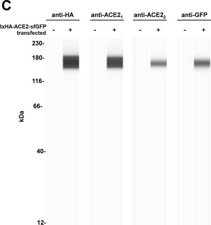
Simple Western: GFP Antibody [Unconjugated] [AF4240] - Simple Western: GFP Antibody [Unconjugated] [AF4240] - Design for a membrane-localized ACE2 expression system. (A) Our ACE2 construct is driven by a CMV promoter followed by the first 25 residues of ACE2 containing the leader sequence that direct ACE2 to the plasma membrane. This is followed by a 3xHA tag linked to the remainder of ACE2 (20-805) and a C-terminal sfGFP. Both 3xHA and sfGFP fusions are separated from ACE2 by flexible 3xGGGGS linkers. (B) The ACE2 fusion protein is designed to be embedded in the plasma membrane where it can perform extracellular carboxypeptidase-mediated metabolism and its levels can be detected by cell staining with antibodies to HA. (C) Lysates from untransfected or 3xHA-ACE2-sfGFP-transfected HEK293 cells were analyzed by automated Jess capillary immunoassay using antibodies to HA, GFP, and two ACE2 antibodies. (D) Confocal fluorescence microscopy of HEK cells transfected with 3xHA-ACE2-sfGFP and stained with HA and the nuclear stain DAPI. Image collected and cropped by CiteAb from the following publication (https://pubmed.ncbi.nlm.nih.gov/37644110), licensed under a CC-BY license. Not internally tested by R&D Systems.
Reconstitution Calculator
Preparation and Storage
- 12 months from date of receipt, -20 to -70 °C as supplied.
- 1 month, 2 to 8 °C under sterile conditions after reconstitution.
- 6 months, -20 to -70 °C under sterile conditions after reconstitution.
Background: GFP
Green fluorescent protein (GFP) is a 27 kDa protein originally isolated from the jellyfish Aequorea victoria. In the presence of UV light (490-520 nm), it emits a green fluorescent color that can be used to pinpoint locations of various intracellular proteins. GFP is 238 amino acids (aa) in length. It is a globular monomer that has a tendency to dimerize. The monomer has the shape of a beta -barrel with a chromophore (aa 65-67) containing alpha -helix running up its center. GFPuv is the Aequorea sequence with three aa substitutions; Phe to Ser at # 99, Met to Thr at # 153, and Val to Ala at # 163. This form expresses faster and is 18-fold brighter than native GFP; excitation peaks at 395 nm and emission at 508 nm.
Product Datasheets
Citations for GFP Antibody
R&D Systems personnel manually curate a database that contains references using R&D Systems products. The data collected includes not only links to publications in PubMed, but also provides information about sample types, species, and experimental conditions.
17
Citations: Showing 1 - 10
Filter your results:
Filter by:
-
Smooth Muscle Cell Reprogramming in Aortic Aneurysms
Authors: Chen PY, Qin L, Li G et al.
Cell Stem Cell
-
A cell-based assay for rapid assessment of ACE2 catalytic function
Authors: Warren M. Meyers, Ryan J. Hong, Wun Chey Sin, Christine S. Kim, Kurt Haas
Scientific Reports
-
Proteomics uncover EPHA2 as a potential novel therapeutic target in colorectal cancer cell lines with acquired cetuximab resistance
Authors: Lucien Torlot, Anna Jarzab, Johanna Albert, Ágnes Pók-Udvari, Arndt Stahler, Julian Walter Holch et al.
Journal of Cancer Research and Clinical Oncology
-
Blood-brain barrier penetration of non-replicating SARS-CoV-2 and S1 Variants of Concern induce neuroinflammation which is accentuated in a mouse model of Alzheimer's disease
Authors: MA Erickson, AF Logsdon, EM Rhea, KM Hansen, SJ Holden, WA Banks, JL Smith, C German, SA Farr, JE Morley, RR Weaver, AJ Hirsch, A Kovac, E Kontsekova, KK Baumann, MA Omer, J Raber
Brain, Behavior, and Immunity, 2023-01-20;0(0):.
Species: Mouse
Sample Types: Whole Tissue
Applications: IHC -
Extending the dynamic range of biomarker quantification through molecular equalization
Authors: Newman, S;Wilson, B;Mamerow, D;Wollant, B;Nyein, H;Rosenberg-Hasson, Y;Maecker, H;Eisenstein, M;Soh, H;
bioRxiv
Species: Human
Sample Types: Serum
Applications: Bioassay -
Intestinal inflammation and increased intestinal permeability in Plasmodium chabaudi AS infected mice
Authors: Jason P Mooney, Sophia M DonVito, Rivka Lim, Marianne Keith, Lia Pickles, Eleanor A Maguire et al.
Wellcome Open Research
-
ESCRT-I fuels lysosomal degradation to restrict TFEB/TFE3 signaling via the Rag-mTORC1 pathway
Authors: Marta Wróbel, Jarosław Cendrowski, Ewelina Szymańska, Malwina Grębowicz-Maciukiewicz, Noga Budick-Harmelin, Matylda Macias et al.
Life Science Alliance
-
MSC Pretreatment for Improved Transplantation Viability Results in Improved Ventricular Function in Infarcted Hearts
Authors: MF Pittenger, S Eghtesad, PG Sanchez, X Liu, Z Wu, L Chen, BP Griffith
International Journal of Molecular Sciences, 2022-01-08;23(2):.
Species: Rat
Sample Types: Cell Lysates
Applications: Western Blot -
Evidence of a Myenteric Plexus Barrier and Its Macrophage-Dependent Degradation During Murine Colitis: Implications in Enteric Neuroinflammation
Authors: D Dora, S Ferenczi, R Stavely, VE Toth, ZV Varga, T Kovacs, I Bodi, R Hotta, KJ Kovacs, AM Goldstein, N Nagy
Cellular and Molecular Gastroenterology and Hepatology, 2021-07-08;0(0):.
Species: Mouse
Sample Types: Whole Tissue
Applications: IHC -
Neonatal diabetes mutations disrupt a chromatin pioneering function that activates the human insulin gene
Authors: I Akerman, MA Maestro, E De Franco, V Grau, S Flanagan, J García-Hur, G Mittler, P Ravassard, L Piemonti, S Ellard, AT Hattersley, J Ferrer
Cell Reports, 2021-04-13;35(2):108981.
Species: Human
Sample Types: Whole Tissue
Applications: IHC -
Wnt/Beta-catenin/Esrrb signalling controls the tissue-scale reorganization and maintenance of the pluripotent lineage during murine embryonic diapause
Authors: R Fan, YS Kim, J Wu, R Chen, D Zeuschner, K Mildner, K Adachi, G Wu, S Galatidou, J Li, HR Schöler, SA Leidel, I Bedzhov
Nat Commun, 2020-10-30;11(1):5499.
Species: Mouse
Sample Types: Whole Cells, Whole Tissue
Applications: ICC, IHC -
Molecular Motor KIF3B Acts as a Key Regulator of Dendritic Architecture in Cortical Neurons
Authors: Nadine F. Joseph, Eddie Grinman, Supriya Swarnkar, Sathyanarayanan V. Puthanveettil
Frontiers in Cellular Neuroscience
-
Splicing variation of BMP2K balances abundance of COPII assemblies and autophagic degradation in erythroid cells
Authors: J Cendrowski, M Kaczmarek, M Mazur, K Kuzmicz-Ko, K Jastrzebsk, M Brewinska-, A Kominek, K Piwocka, M Miaczynska
Elife, 2020-08-14;9(0):.
Species: Human
Sample Types: Cell Lysates, Whole Cells
Applications: Co-IP, ICC, Western Blot -
Agrin has a pathological role in the progression of oral cancer
Authors: C Rivera, FS Zandonadi, C Sánchez-Ro, CD Soares, DC Granato, WA González-A, AF Paes Leme
Br. J. Cancer, 2018-06-06;0(0):.
Applications: Immunoprecipitation -
Natural killer cells attenuate cytomegalovirus-induced hearing loss in mice
Authors: Ali A. Almishaal, Pranav D. Mathur, Elaine Hillas, Liting Chen, Anne Zhang, Jun Yang et al.
PLOS Pathogens
-
Lymphatic endothelial progenitors originate from plastic myeloid cells activated by toll-like receptor-4
Authors: LD Volk-Drape, KL Hall, AC Wilber, S Ran
PLoS ONE, 2017-06-09;12(6):e0179257.
Species: Mouse
Sample Types: Whole Tissue
Applications: IHC -
Efficient Generation of Cardiac Purkinje Cells from ESCs by Activating cAMP Signaling
Authors: Su-Yi Tsai, Karen Maass, Jia Lu, Glenn I. Fishman, Shuibing Chen, Todd Evans
Stem Cell Reports
FAQs
No product specific FAQs exist for this product, however you may
View all Antibody FAQsReviews for GFP Antibody
Average Rating: 4 (Based on 3 Reviews)
Have you used GFP Antibody?
Submit a review and receive an Amazon gift card.
$25/€18/£15/$25CAN/¥75 Yuan/¥2500 Yen for a review with an image
$10/€7/£6/$10 CAD/¥70 Yuan/¥1110 Yen for a review without an image
Filter by:


