Human Akt1 (E17K Mutation) Antibody Summary
Accession # P31749
Customers also Viewed
Applications
Please Note: Optimal dilutions should be determined by each laboratory for each application. General Protocols are available in the Technical Information section on our website.
Scientific Data
 View Larger
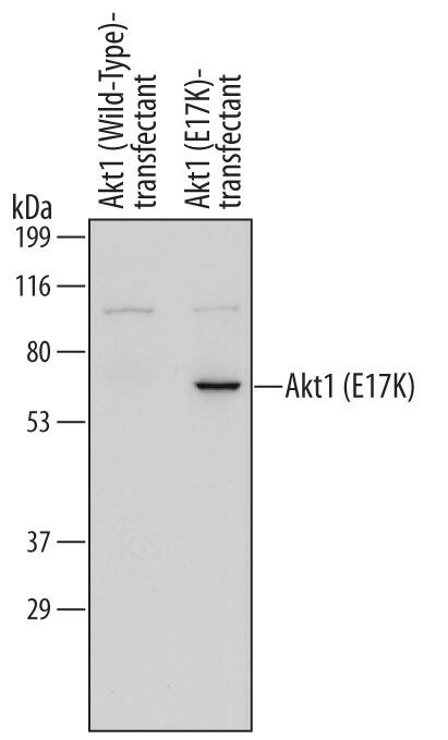
Detection of Human Akt1 (E17K Mutation) by Western Blot. Western blot shows lysates of 293-EBNA human EBV-expressing embryonic kidney cell line either transfected with Akt1 (wild-type) or transfected with Akt1 (E17K Mutation). PVDF membrane was probed with 0.5 µg/mL of Mouse Anti-Human Akt1 (E17K Mutation) Monoclonal Antibody (Catalog # MAB6815) followed by HRP-conjugated Anti-Mouse IgG Secondary Antibody (Catalog # HAF007). A specific band was detected for Akt1 (E17K Mutation) at approximately 60 kDa (as indicated). This experiment was conducted under reducing conditions and using Immunoblot Buffer Group 1.
 View Larger
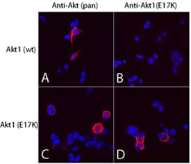
Akt1 (E17K Mutation) in 293T Human Cell Line. Akt pan specific (panels A and C) and Akt1 (E17K Mutation) (panels B and D) were detected in immersion fixed 293T human embryonic kidney cell line transfected with wild type Akt1 (panels A and B) or E17K mutated Akt1 (panels C and D) using Mouse Anti-Human Akt1 (E17K Mutation) Monoclonal Antibody (Catalog # MAB6815) and Mouse Anti-Human/Mouse/Rat Akt Pan Specific Monoclonal Antibody (Catalog # MAB2055). Both antibodies were used at 10 µg/mL for 3 hours at room temperature. Cells were stained using the NorthernLights™ 557-conjugated Anti-Mouse IgG Secondary Antibody (red; Catalog # NL007) and counterstained with DAPI (blue). Specific staining was localized to plasma membranes and cytoplasm. View our protocol for Fluorescent ICC Staining of Cells on Coverslips.
Preparation and Storage
- 12 months from date of receipt, -20 to -70 °C as supplied.
- 1 month, 2 to 8 °C under sterile conditions after reconstitution.
- 6 months, -20 to -70 °C under sterile conditions after reconstitution.
Background: Akt1
Akt, also known as protein kinase B (PKB), is a central kinase in such diverse cellular processes as glucose uptake, cell cycle progression, and apoptosis. Three highly homologous members define the Akt family: Akt1 (PKB alpha ), Akt2 (PKB beta ), and Akt3 (PKB gamma ). All three Akts contain an amino-terminal pleckstrin homology domain, a central kinase domain, and a carboxyl-terminal regulatory domain. Akt1 is the most widely expressed family member and is frequently activated in a number of carcinomas, including breast, prostate, lung, pancreatic, liver, ovarian, and colorectal cancer. Akt1 is activated in a multistep process that involves the sequential phosphorylation of Thr450 by JNK kinases, Thr308 by PDK1, and Ser473 by PDK2 or mTORC2. Activated Akt1 phosphorylates a wide variety of cytosolic, nuclear, and mitochondrial substrates. Substitution of glutamic acid with lysine at amino acid 17 (E17K) occurs in multiple malignancies and results in constitutive Akt1 activation and cell transformation. Within aa 5‑30, human Akt1 shares 100% aa sequence identity with mouse and rat Akt1.
- Carpten, J.D. et al. (2007) Nature 448:439.
- Bleeker, F.E. et al. (2008) Oncogene 27:5648.
- Brugge, J. et al. Cancer Cell 12:104.
Product Datasheets
FAQs
No product specific FAQs exist for this product, however you may
View all Antibody FAQsReviews for Human Akt1 (E17K Mutation) Antibody
There are currently no reviews for this product. Be the first to review Human Akt1 (E17K Mutation) Antibody and earn rewards!
Have you used Human Akt1 (E17K Mutation) Antibody?
Submit a review and receive an Amazon gift card.
$25/€18/£15/$25CAN/¥75 Yuan/¥2500 Yen for a review with an image
$10/€7/£6/$10 CAD/¥70 Yuan/¥1110 Yen for a review without an image


