Human ALCAM/CD166 PE-conjugated Antibody Summary
Trp28-Ala526
Accession # Q13740
Customers also Viewed
Applications
Please Note: Optimal dilutions should be determined by each laboratory for each application. General Protocols are available in the Technical Information section on our website.
Scientific Data
 View Larger
Detection of ALCAM/CD166 in Human Whole Blood Monocytes by Flow Cytometry. Human whole blood monocytes were stained with Mouse Anti-Human ALCAM/CD166 PE-conjugated Monoclonal Antibody (Catalog # FAB6561P, filled histogram) or isotype control antibody (IC002P, open histogram). View our protocol for Staining Membrane-associated Proteins.
 View Larger
Detection of Human ALCAM/CD166 by Flow Cytometry Analysis of CSC marker expression in TOP-GFP cultures.(A) Representative images of the three independent single-cell-cloned CSC cultures, lentivirally transduced with TOP-GFP. Phase contrast (top) and fluorescence microscopy (bottom) for each of the cultures indicated. Bar = 90 µm. (B) Single parameter histograms for GFP intensity for each of the TOP-GFP single-cell-cloned CSC cultures with the TOP-GFPlow (10% lowest) and TOP-GFPhigh (10% highest) populations indicated. (C) Single parameter histograms for the indicated cell surface markers for each of the indicated cultures. Gray denotes TOP-GFPlow (10% lowest) and green denotes TOP-GFPhigh (10% highest) populations. (D) Density plots for CD29/CD24 and CD44/CD166 from TOP-GFPlow (gray) and TOP-GFPhigh (green) populations of each culture. Additional details for this experiment can be found at https://osf.io/tfy28/.Flow cytometry gating strategy.Representative density plots of gating strategy to assess cell surface markers from TOP-GFPlow and TOP-GFPhigh populations. Forward scatter area (FSC-A) and PerCP-Cy5.5 was used to gate on viable cells (PI negative cells), followed by forward verses side scatter area (FSC-A vs SSC-A) to identify cells of interest and exclude debris, which were then analyzed by FSC-A and forward scatter width (FSC-W), and then SSC-A and side scatter width (SSC-W) to exclude doublet cells. From the single-cell population, SSC and FITC were used to gate on the TOP-GFPlow (10% lowest) and TOP-GFPhigh (10% highest) populations. TOP-GFPlow and TOP-GFPhigh populations were then assessed for PE and APC to detect the fluorophores conjugated to antibodies against the cell surface markers analyzed in this study. Additional details for this experiment can be found at https://osf.io/tfy28/. Image collected and cropped by CiteAb from the following publication (https://pubmed.ncbi.nlm.nih.gov/31215867), licensed under a CC-BY license. Not internally tested by R&D Systems.
 View Larger
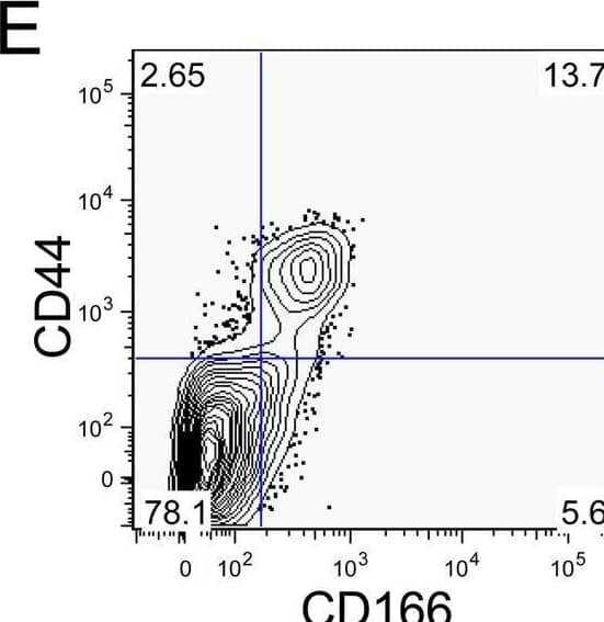
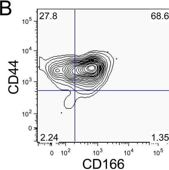
Detection of ALCAM/CD166 by Flow Cytometry In vitro maintenance and expansion of CoCSC.Human ESA+CD44+CD166+ cells were plated in limiting dilution and cultured for fourteen days in serum-free maintenance conditions. Colorectal tumor colonies were then either analyzed for A) ESA expression by IHC, or B) ESA, CD44 and CD166 expression by flow cytometry. C) Cellular phenotype of single colony-derived tumors, showing human ESA+ cell subpopulations expressing CD44 and CD166. D) Tumor growth curves are shown for either 2,000 CoCSC phenotype cells or an equal number of cells with all other phenotypes (Other), which were isolated from in vitro colony-derived tumors (tumors/animals injected). Inset shows lentiviral insertion band obtained by inverse PCR of 1) human xenograft tumor cells, 2) ESA+CD44+CD166+ (CoCSC) cells or 3) ESA+CD44− (Other) cells isolated by FACS. Phenotypic and morphological analysis of single-cell derived tumors from serially transplanted CoCSC show that the diverse E) phenotype and F) histological makeup of xenogeneic colorectal tumors are maintained following brief in vitro culture in limiting dilution. Black bar = 100 µm. Image collected and cropped by CiteAb from the following open publication (https://pubmed.ncbi.nlm.nih.gov/18560594), licensed under a CC-BY license. Not internally tested by R&D Systems.
Preparation and Storage
Background: ALCAM/CD166
ALCAM, activated leukocyte cell adhesion molecule, is a type I membrane glycoprotein and a member of the immunoglobulin supergene family. It is also known as CD166, MEMD, SC-1/DM-GRASP/BEN in the chicken, and KG-CAM in the rat. ALCAM is expressed on thymic epithelial cells, activated B and T cells, and monocytes. ALCAM can bind itself homotypically and is also capable of binding CD6, NgCAM, and other, as of yet, unidentified brain proteins. The ALCAM/CD6 interaction may be involved in T cell development and T cell regulation. Additionally, ALCAM/CD6 and ALCAM/NgCAM interactions may play roles in the nervous system. ALCAM has also been observed to be upregulated on highly metastasizing melanoma cell lines and may play a role in tumor migration. ALCAM is a 583 amino acid (aa) protein consisting of a 27 aa signal peptide, a 500 aa extracellular domain, a 24 aa transmembrane domain and a 32 aa cytoplasmic domain. The extracellular domain of ALCAM contains 5 Ig-like domains.
- Bowen, M.A. et al. (1995) J. Exp. Med. 181:2213.
- Aruffo, A. et al. (1997) Immunol. Today 18:498.
- Degen, W.G. et al. (1998) Am. J. Pathol. 152:805.
Product Datasheets
Citations for Human ALCAM/CD166 PE-conjugated Antibody
R&D Systems personnel manually curate a database that contains references using R&D Systems products. The data collected includes not only links to publications in PubMed, but also provides information about sample types, species, and experimental conditions.
17
Citations: Showing 1 - 10
Filter your results:
Filter by:
-
Influence of glycan structure on the colonization of Streptococcus pneumoniae on human respiratory epithelial cells
Authors: YY Chun, KS Tan, L Yu, M Pang, MHM Wong, R Nakamoto, WZ Chua, A Huee-Ping, ZZR Lew, HH Ong, VT Chow, T Tran, D Yun Wang, LT Sham
Proceedings of the National Academy of Sciences of the United States of America, 2023-03-21;120(13):e2213584120.
-
B4GALNT1 induces angiogenesis, anchorage independence growth and motility, and promotes tumorigenesis in melanoma by induction of ganglioside GM2/GD2
Authors: H Yoshida, L Koodie, K Jacobsen, K Hanzawa, Y Miyamoto, M Yamamoto
Sci Rep, 2020-01-27;10(1):1199.
Species: Human
Sample Types: Whole Cells
Applications: Flow Cytometry -
Physiologic expansion of human heart-derived cells enhances therapeutic repair of injured myocardium
Authors: S Mount, P Kanda, S Parent, S Khan, C Michie, L Davila, V Chan, RA Davies, H Haddad, D Courtman, DJ Stewart, DR Davis
Stem Cell Res Ther, 2019-11-04;10(1):316.
Species: Human
Sample Types: Whole Cells
Applications: Flow Cytometry -
Replication Study: Wnt activity defines colon cancer stem cells and is regulated by the microenvironment
Authors: Anthony Essex, Javier Pineda, Grishma Acharya, Hong Xin, James Evans, Elizabeth Iorns et al.
eLife
-
Methionine is a metabolic dependency of tumor-initiating cells
Authors: Z Wang, LY Yip, JHJ Lee, Z Wu, HY Chew, PKW Chong, CC Teo, HY Ang, KLE Peh, J Yuan, S Ma, LSK Choo, N Basri, X Jiang, Q Yu, AM Hillmer, WT Lim, TKH Lim, A Takano, EH Tan, DSW Tan, YS Ho, B Lim, WL Tam
Nat. Med., 2019-05-06;25(5):825-837.
Species: Human
Sample Types: Whole Cells
Applications: Flow Cytometry -
Reduced CD146 expression promotes tumorigenesis and cancer stemness in colorectal cancer through activating Wnt/beta-catenin signaling.
Authors: Liu D, DU L, Chen D, Ye Z, Duan H, Tu T, Feng J, Yang Y, Chen Q, Yan X
Oncotarget, 2016-06-28;7(26):40704-40718.
Species: Human
Sample Types: Whole Cells
Applications: Flow Cytometry -
Registered report: Wnt activity defines colon cancer stem cells and is regulated by the microenvironment
Authors: James Evans, Anthony Essex, Hong Xin, Nurith Amitai, Lindsey Brinton, Erin Griner et al.
eLife
-
CD166/ALCAM expression is characteristic of tumorigenicity and invasive and migratory activities of pancreatic cancer cells.
Authors: Fujiwara K, Ohuchida K, Sada M, Horioka K, Ulrich C, Shindo K, Ohtsuka T, Takahata S, Mizumoto K, Oda Y, Tanaka M
PLoS ONE, 2014-09-15;9(9):e107247.
Species: Human
Sample Types: Whole Cells
Applications: Flow Cytometry -
Establishment of highly tumorigenic human colorectal cancer cell line (CR4) with properties of putative cancer stem cells.
Authors: Rowehl, Rebecca, Burke, Stephani, Bialkowska, Agnieszk, Pettet, Donald W, Rowehl, Leahana, Li, Ellen, Antoniou, Eric, Zhang, Yuanhao, Bergamaschi, Roberto, Shroyer, Kenneth, Ojima, Iwao, Botchkina, Galina I
PLoS ONE, 2014-06-12;9(6):e99091.
Species: Human
Sample Types: Whole Cells
Applications: Flow Cytometry -
Activated leukocyte cell adhesion molecule (CD166): an "inert" cancer stem cell marker for non-small cell lung cancer?
Authors: Tachezy M, Zander H, Wolters-Eisfeld G, Muller J, Wicklein D, Gebauer F, Izbicki J, Bockhorn M
Stem Cells, 2014-06-01;32(6):1429-36.
Species: Human
Sample Types: Whole Cells
Applications: Flow Cytometry -
Phenotyping of human melanoma cells reveals a unique composition of receptor targets and a subpopulation co-expressing ErbB4, EPO-R and NGF-R.
Authors: Mirkina I, Hadzijusufovic E, Krepler C, Mikula M, Mechtcheriakova D, Strommer S, Stella A, Jensen-Jarolim E, Holler C, Wacheck V, Pehamberger H, Valent P
PLoS ONE, 2014-01-29;9(1):e84417.
Species: Human
Sample Types: Whole Cells
Applications: Flow Cytometry -
Cultivation and characterization of cornea limbal epithelial stem cells on lens capsule in animal material-free medium.
Authors: Albert R, Vereb Z, Csomos K, Moe M, Johnsen E, Olstad O, Nicolaissen B, Rajnavolgyi E, Fesus L, Berta A, Petrovski G
PLoS ONE, 2012-10-09;7(10):e47187.
Species: Human
Sample Types: Whole Cells
Applications: Flow Cytometry -
CD166(pos) subpopulation from differentiated human ES and iPS cells support repair of acute lung injury.
Authors: Soh, Boon Sen, Zheng, Dahai, Li Yeo, Julie Su, Yang, Henry He, Ng, Shi Yan, Wong, Lan Hion, Zhang, Wencai, Li, Pin, Nichane, Massimo, Asmat, Atasha, Wong, Poo Sing, Wong, Peng Che, Su, Lin Lin, Mantalaris, Sakis A, Lu, Jia, Xian, Wa, McKeon, Frank, Chen, Jianzhu, Lim, Elaine H, Lim, Bing
Mol Ther, 2012-09-11;20(12):2335-46.
Species: Human
Sample Types: Whole Cells
Applications: Cell Selection, Flow Cytometry -
Epstein-Barr virus latent membrane protein 1 induces cancer stem/progenitor-like cells in nasopharyngeal epithelial cell lines.
Authors: Kondo S, Wakisaka N, Muramatsu M
J. Virol., 2011-08-17;85(21):11255-64.
Species: Human
Sample Types: Whole Cells
Applications: Flow Cytometry -
Long-lasting inhibitory effects of fetal liver mesenchymal stem cells on T-lymphocyte proliferation.
Authors: Giuliani M, Fleury M, Vernochet A, Ketroussi F, Clay D, Azzarone B, Lataillade JJ, Durrbach A
2011-05-19;6(5):e19988.
Species: Human
Sample Types: Whole Cells
Applications: Flow Cytometry -
Multipotent mesenchymal progenitor cells are present in endarterectomized tissues from patients with chronic thromboembolic pulmonary hypertension.
Authors: Firth AL, Yao W, Ogawa A, Madani MM, Lin GY, Yuan JX
Am. J. Physiol., Cell Physiol., 2010-02-24;298(5):C1217-25.
Species: Human
Sample Types: Whole Cells
Applications: Flow Cytometry, ICC -
Substantial differences between human and ovine mesenchymal stem cells in response to osteogenic media: how to explain and how to manage?
Authors: Kalaszczynska Ilona, Ruminski Slawomir, Platek Anna E et al.
BioResearch Open Access
FAQs
No product specific FAQs exist for this product, however you may
View all Antibody FAQsReviews for Human ALCAM/CD166 PE-conjugated Antibody
There are currently no reviews for this product. Be the first to review Human ALCAM/CD166 PE-conjugated Antibody and earn rewards!
Have you used Human ALCAM/CD166 PE-conjugated Antibody?
Submit a review and receive an Amazon gift card.
$25/€18/£15/$25CAN/¥75 Yuan/¥2500 Yen for a review with an image
$10/€7/£6/$10 CAD/¥70 Yuan/¥1110 Yen for a review without an image


