Human C10orf99 Antibody Summary
Applications
This antibody functions as an ELISA detection antibody when paired with Mouse Anti-Human C10orf99 Monoclonal Antibody (Catalog # MAB10416).
This product is intended for assay development on various assay platforms requiring antibody pairs.
Please Note: Optimal dilutions should be determined by each laboratory for each application. General Protocols are available in the Technical Information section on our website.
Scientific Data
 View Larger
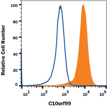
Detection of C10orf99 in SW480 human cell line. SW480 human cell line was stained with Mouse Anti-Human C10orf99 Monoclonal Antibody (Catalog # MAB104161, filled histogram) or isotype control antibody (MAB0041, open histogram), followed by APC-conjugated Anti-Mouse IgG Secondary Antibody (F0101B). To facilitate intracellular staining, cells were fixed with Flow Cytometry Fixation Buffer (FC004) and permeabilized with Flow Cytometry Permeabilization/Wash Buffer I (FC005). Staining was performed using our Staining Intracellular Molecules protocol.
 View Larger
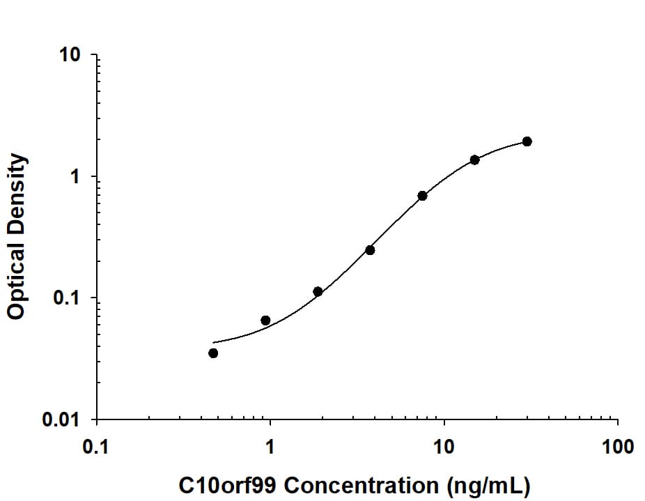
Human C10orf99 ELISA Standard Curve. Recombinant Human C10orf99 protein was serially diluted 2-fold and captured by Mouse Anti-Human C10orf99 Monoclonal Antibody (Catalog # MAB10416) coated on a Clear Polystyrene Microplate (DY990). Mouse Anti-Human C10orf99 Monoclonal Antibody (Catalog # MAB104161) was biotinylated and incubated with the protein captured on the plate. Detection of the standard curve was achieved by incubating Streptavidin-HRP (DY998) followed by Substrate Solution (DY999) and stopping the enzymatic reaction with Stop Solution (DY994).
 View Larger
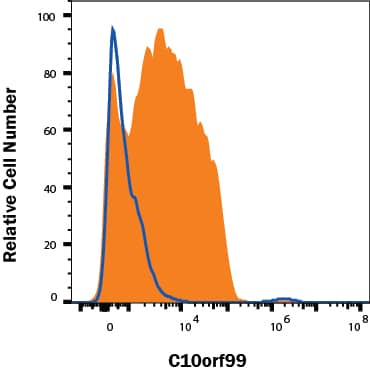
Detection of C10orf99 in T84 cells by Flow Cytometry. T84 human epithelial carcinoma cells treated with 5 μg/ml Brefeldin A 1231/5 for 5 hours were stained with Mouse Anti-Human C10orf99 Monoclonal Antibody (Catalog # MAB104161, filled histogram) or isotype control antibody (Catalog # MAB0041, open histogram), followed by Allophycocyanin-conjugated Anti-Mouse IgG Secondary Antibody (Catalog # F0101B). To facilitate intracellular staining, cells were fixed with Flow Cytometry Fixation Buffer (Catalog # FC004) and permeabilized with Flow Cytometry Permeabilization/Wash Buffer I (Catalog # FC005). View our protocol for Staining Intracellular Molecules.
Reconstitution Calculator
Preparation and Storage
- 12 months from date of receipt, -20 to -70 °C as supplied.
- 1 month, 2 to 8 °C under sterile conditions after reconstitution.
- 6 months, -20 to -70 °C under sterile conditions after reconstitution.
Background: C10orf99
Human Chromosome 10 open reading frame 99 is a 9.2 KDa protein encoded by the C10orf99 gene. C10orf99 has been described as a potential cytokine and a ligand Sushi Domain Containing 2 (SUSD2), inducing SUSD2 internalization upon binding. It has also been proposed to be a chemotactic factor that mediates lymphocytes recruitment to epithelia through binding and activation of the G-protein coupled receptor GPR15.
Product Datasheets
FAQs
No product specific FAQs exist for this product, however you may
View all Antibody FAQsReviews for Human C10orf99 Antibody
There are currently no reviews for this product. Be the first to review Human C10orf99 Antibody and earn rewards!
Have you used Human C10orf99 Antibody?
Submit a review and receive an Amazon gift card.
$25/€18/£15/$25CAN/¥75 Yuan/¥2500 Yen for a review with an image
$10/€7/£6/$10 CAD/¥70 Yuan/¥1110 Yen for a review without an image

