Human CD4 Alexa Fluor® 488-conjugated Antibody Summary
Lys26-Trp390
Accession # P01730
Customers also Viewed
Applications
Please Note: Optimal dilutions should be determined by each laboratory for each application. General Protocols are available in the Technical Information section on our website.
Scientific Data
 View Larger
Detection of CD4 in Human Blood Lymphocytes by Flow Cytometry. Human peripheral blood lymphocytes were stained with Goat Anti-Human CD4 Alexa Fluor® 488-conjugated Antigen Affinity-purified Polyclonal Antibody (Catalog # FAB8165G, filled histogram) or isotype control antibody (Catalog # IC108G, open histogram). View our protocol for Staining Membrane-associated Proteins.
 View Larger
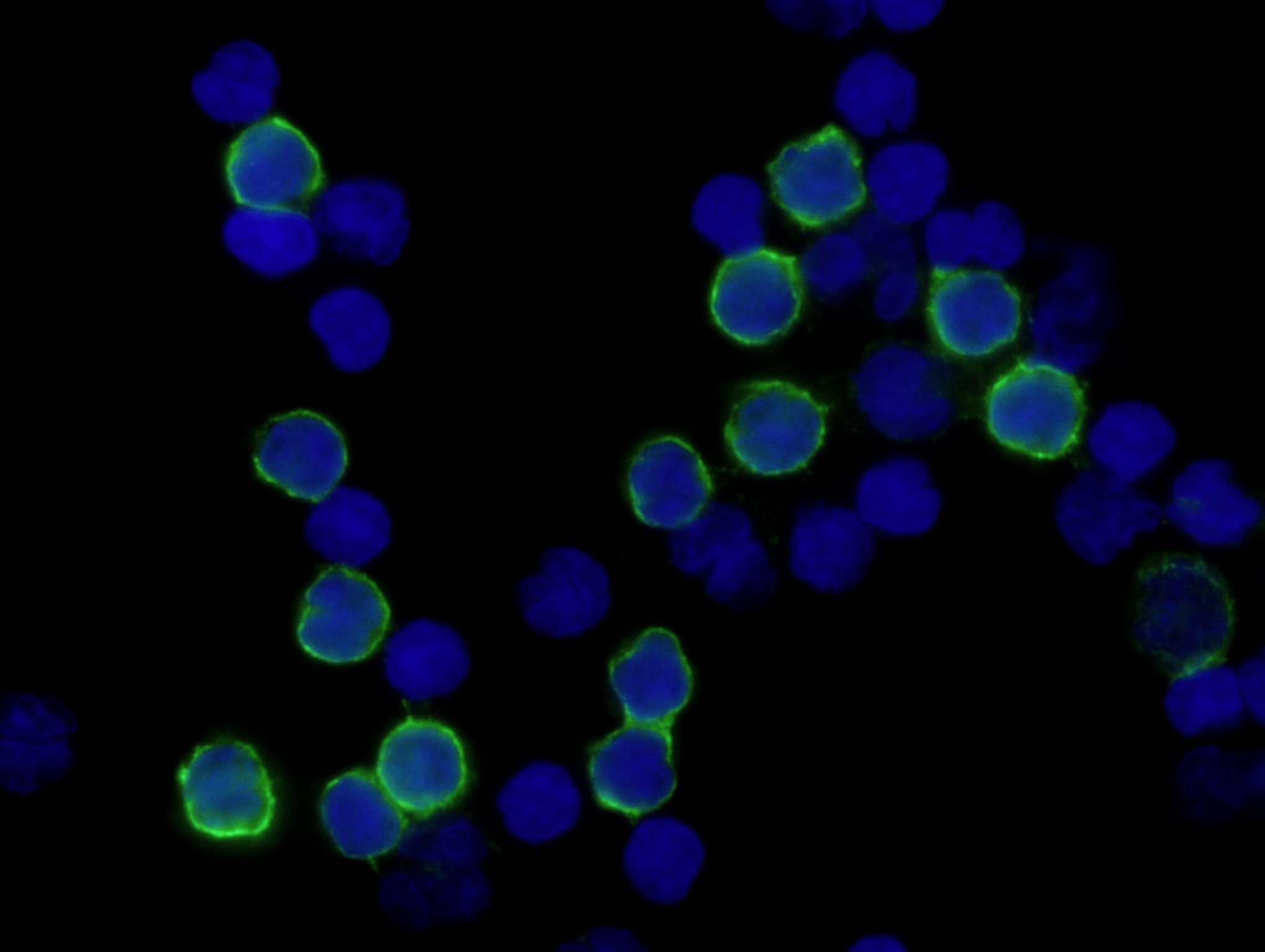
CD4 in Human PBMCs. CD4 was detected in immersion fixed human peripheral blood mononuclear cells (PBMCs) using Goat Anti-Human CD4 Alexa Fluor® 488-conjugated Antigen Affinity-purified Polyclonal Antibody (Catalog # FAB8165G, green) at 10 µg/mL for 3 hours at room temperature. Cells were counterstained with DAPI (blue). Specific staining was localized to cell surfaces. View our protocol for Fluorescent ICC Staining of Non-adherent Cells.
Preparation and Storage
- 12 months from date of receipt, 2 to 8 °C as supplied.
Background: CD4
CD4 is an approximately 55 kDa type I membrane glycoprotein that is expressed predominantly on most thymocytes and a subset of mature T lymphocytes. In humans, CD4 is also expressed to a lesser extent on monocytes and macrophage related cells. Human CD4 cDNA encodes a 458 amino acid (aa) residue precursor protein with a 25 aa residue signal peptide, a 371 aa residue extracellular region containing four immunoglobulin homology domains, a 24 aa residue transmembrane domain and a 38 aa residue cytoplasmic domain. CD4 is a coreceptor required for T cell recognition of antigens that are presented by class II major histocompatibility complexes. CD4 has been shown to be a coreceptor of HIV entry and specifically binds gp120, the external envelope glycoprotein of HIV.
Product Datasheets
Product Specific Notices
This product is provided under an agreement between Life Technologies Corporation and R&D Systems, Inc, and the manufacture, use, sale or import of this product is subject to one or more US patents and corresponding non-US equivalents, owned by Life Technologies Corporation and its affiliates. The purchase of this product conveys to the buyer the non-transferable right to use the purchased amount of the product and components of the product only in research conducted by the buyer (whether the buyer is an academic or for-profit entity). The sale of this product is expressly conditioned on the buyer not using the product or its components (1) in manufacturing; (2) to provide a service, information, or data to an unaffiliated third party for payment; (3) for therapeutic, diagnostic or prophylactic purposes; (4) to resell, sell, or otherwise transfer this product or its components to any third party, or for any other commercial purpose. Life Technologies Corporation will not assert a claim against the buyer of the infringement of the above patents based on the manufacture, use or sale of a commercial product developed in research by the buyer in which this product or its components was employed, provided that neither this product nor any of its components was used in the manufacture of such product. For information on purchasing a license to this product for purposes other than research, contact Life Technologies Corporation, Cell Analysis Business Unit, Business Development, 29851 Willow Creek Road, Eugene, OR 97402, Tel: (541) 465-8300. Fax: (541) 335-0354.
Citations for Human CD4 Alexa Fluor® 488-conjugated Antibody
R&D Systems personnel manually curate a database that contains references using R&D Systems products. The data collected includes not only links to publications in PubMed, but also provides information about sample types, species, and experimental conditions.
13
Citations: Showing 1 - 10
Filter your results:
Filter by:
-
Targeting Pin1 renders pancreatic cancer eradicable by synergizing with immunochemotherapy
Authors: Koikawa K, Kibe S, Suizu F Et al.
Cell
-
Highly multiplexed immunofluorescence imaging of human tissues and tumors using t-CyCIF and conventional optical microscopes.
Authors: Lin J. R, Izar B, et al.
Elife
-
Multiplexed imaging and automated signal quantification in formalin-fixed paraffin-embedded tissues by ChipCytometry
Authors: Jarosch S, KOhlen J, Sarker R Et al.
Cell Rep Methods
-
Spatial intra-tumor heterogeneity is associated with survival of lung adenocarcinoma patients
Authors: Hua-Jun Wu, Daniel Temko, Zoltan Maliga, Andre L. Moreira, Emi Sei, Darlan Conterno Minussi et al.
Cell Genomics
-
ChipCytometry for multiplexed detection of protein and mRNA markers on human FFPE tissue samples
Authors: Sebastian Jarosch, Jan Köhlen, Sabrina Wagner, Elvira D’Ippolito, Dirk H. Busch
STAR Protocols
-
Potent anti-viral activity of a trispecific HIV neutralizing antibody in SHIV-infected monkeys
Authors: A Pegu, L Xu, ME DeMouth, G Fabozzi, K March, CG Almasri, MD Cully, K Wang, ES Yang, J Dias, CM Fennessey, J Hataye, RR Wei, E Rao, JP Casazza, W Promsote, M Asokan, K McKee, SD Schmidt, X Chen, C Liu, W Shi, H Geng, KE Foulds, SF Kao, A Noe, H Li, GM Shaw, T Zhou, C Petrovas, JP Todd, BF Keele, JD Lifson, NA Doria-Rose, RA Koup, ZY Yang, GJ Nabel, JR Mascola
Cell Reports, 2022-01-04;38(1):110199.
Species: Rhesus Macaque
Sample Types: Whole Tissue
Applications: IHC -
Antigen dominance hierarchies shape TCF1+ progenitor CD8 T cell phenotypes in tumors
Authors: Megan L. Burger, Amanda M. Cruz, Grace E. Crossland, Giorgio Gaglia, Cecily C. Ritch, Sarah E. Blatt et al.
Cell
-
Targeting immunosuppressive macrophages overcomes PARP inhibitor resistance in BRCA1-associated triple-negative breast cancer
Authors: Anita K. Mehta, Emily M. Cheney, Christina A. Hartl, Constantia Pantelidou, Madisson Oliwa, Jessica A. Castrillon et al.
Nature Cancer
-
Highly multiplexed immunofluorescence images and single-cell data of immune markers in tonsil and lung cancer
Authors: Rumana Rashid, Giorgio Gaglia, Yu-An Chen, Jia-Ren Lin, Ziming Du, Zoltan Maliga et al.
Scientific Data
-
Qualifying antibodies for image-based immune profiling and multiplexed tissue imaging
Authors: Ziming Du, Jia-Ren Lin, Rumana Rashid, Zoltan Maliga, Shu Wang, Jon C. Aster et al.
Nature Protocols
-
Accumulation of follicular CD8+ T cells in pathogenic SIV infection
Authors: S Ferrando-M, E Moysi, A Pegu, S Andrews, K Nganou Mak, D Ambrozak, AB McDermott, D Palesch, M Paiardini, GN Pavlakis, JM Brenchley, D Douek, JR Mascola, C Petrovas, RA Koup
J. Clin. Invest., 2018-04-16;0(0):.
Species: Primate
Sample Types: Whole Cells
Applications: ICC -
Treatment with native heterodimeric IL-15 increases cytotoxic lymphocytes and reduces SHIV RNA in lymph nodes
Authors: DC Watson, E Moysi, A Valentin, C Bergamasch, S Devasundar, SP Fortis, J Bear, E Chertova, J Bess, R Sowder, DJ Venzon, C Deleage, JD Estes, JD Lifson, C Petrovas, BK Felber, GN Pavlakis
PLoS Pathog., 2018-02-23;14(2):e1006902.
Species: Primate - Macaca mulatta (Rhesus Macaque)
Sample Types: Whole Tissue
Applications: Confocal Microscopy -
Quantitative Multiplexed Imaging Analysis Reveals a Strong Association between Immunogen-Specific B Cell Responses and Tonsillar Germinal Center Immune Dynamics in Children after Influenza Vaccination
Authors: D Amodio, N Cotugno, G Macchiarul, S Rocca, Y Dimopoulos, MR Castrucci, R De Vito, FM Tucci, AB McDermott, S Narpala, P Rossi, RA Koup, P Palma, C Petrovas
J. Immunol., 2017-12-13;0(0):.
Species: Human
Sample Types: Whole Tissue
Applications: IHC
FAQs
No product specific FAQs exist for this product, however you may
View all Antibody FAQsReviews for Human CD4 Alexa Fluor® 488-conjugated Antibody
There are currently no reviews for this product. Be the first to review Human CD4 Alexa Fluor® 488-conjugated Antibody and earn rewards!
Have you used Human CD4 Alexa Fluor® 488-conjugated Antibody?
Submit a review and receive an Amazon gift card.
$25/€18/£15/$25CAN/¥75 Yuan/¥2500 Yen for a review with an image
$10/€7/£6/$10 CAD/¥70 Yuan/¥1110 Yen for a review without an image


