Human CHD1 Antibody Summary
Lys1531-Thr1710
Accession # O14646
Applications
Please Note: Optimal dilutions should be determined by each laboratory for each application. General Protocols are available in the Technical Information section on our website.
Scientific Data
 View Larger
Detection of Human and Mouse CHD1 by Western Blot. Western blot shows lysates of Jurkat human acute T cell leukemia cell line, K562 human chronic myelogenous leukemia cell line, Raji human Burkitt's lymphoma cell line, and BaF3 mouse pro-B cell line. PVDF Membrane was probed with 0.5 µg/mL of Human CHD1 Monoclonal Antibody (Catalog # MAB6195) followed by HRP-conjugated Anti-Mouse IgG Secondary Antibody (Catalog # HAF007). A specific band was detected for CHD1 at approximately 220 kDa (as indicated). This experiment was conducted under reducing conditions and using Immunoblot Buffer Group 1.
 View Larger
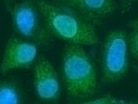
CHD1 in BG01V Human Stem Cells. CHD1 was detected in immersion fixed BG01V human embryonic stem cells using Human CHD1 Monoclonal Antibody (Catalog # MAB6195) at 10 µg/mL for 3 hours at room temperature. Cells were stained using the NorthernLights™ 557-conjugated Anti-Mouse IgG Secondary Antibody (red, upper panel; Catalog # NL007) and counterstained with DAPI (blue, lower panel). Specific staining was localized to nuclei. View our protocol for Fluorescent ICC Staining of Cells on Coverslips.
 View Larger
Western Blot Shows Human CHD1 Specificity by Using Knockout Cell Line. Western blot shows lysates of HEK293T human embryonic kidney parental cell line and CHD1 knockout HEK293T cell line (KO). PVDF membrane was probed with 0.5 µg/mL of Mouse Anti-Human CHD1 Monoclonal Antibody (Catalog # MAB6195) followed by HRP-conjugated Anti-Mouse IgG Secondary Antibody (Catalog # HAF018). A specific band was detected for CHD1 at approximately 210 kDa (as indicated) in the parental HEK293T cell line, but is not detectable in knockout HEK293T cell line. GAPDH (Catalog # MAB5718) is shown as a loading control. This experiment was conducted under reducing conditions and using Immunoblot Buffer Group 1.
Reconstitution Calculator
Preparation and Storage
- 12 months from date of receipt, -20 to -70 °C as supplied.
- 1 month, 2 to 8 °C under sterile conditions after reconstitution.
- 6 months, -20 to -70 °C under sterile conditions after reconstitution.
Background: CHD1
CHD1 (Chromohelicase/ATPase DNA-binding protein 1) is a 200-205 kDa member of the SNF2/RAD54 helicase family of proteins. It is an ATP-dependent chromatin remodeling factor that helps maintain chromatin in a transcriptionally active state. In embryonic stem cells, CHD1 associates with the promoters of active genes, a condition that is associated with open chromatin and pluripotency. Human CHD1 is 1710 amino acids (aa) in length. It contains two Ser-rich regions (aa 1-70 and 117‑137), two Chromo (chromatin-organizer-modifer) domains (aa 272-364 and 389-452), a SNF2 family N-terminal domain (aa 484-763) and a C-terminal helicase domain (aa 792-943). There are at least 30 Ser/Thr phosphorylation sites. Over aa 1531-1710, human CHD1 shares 92% aa identity with mouse CHD1.
Product Datasheets
FAQs
No product specific FAQs exist for this product, however you may
View all Antibody FAQsReviews for Human CHD1 Antibody
Average Rating: 5 (Based on 1 Review)
Have you used Human CHD1 Antibody?
Submit a review and receive an Amazon gift card.
$25/€18/£15/$25CAN/¥75 Yuan/¥2500 Yen for a review with an image
$10/€7/£6/$10 CAD/¥70 Yuan/¥1110 Yen for a review without an image
Filter by:


