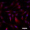
Human Complement Component C1s Antibody Summary
2% cross‑reactivity with recombinant human C1r is observed.
Glu16-Asp688
Accession # P09871
Applications
Please Note: Optimal dilutions should be determined by each laboratory for each application. General Protocols are available in the Technical Information section on our website.
Reconstitution Calculator
Preparation and Storage
- 12 months from date of receipt, -20 to -70 °C as supplied.
- 1 month, 2 to 8 °C under sterile conditions after reconstitution.
- 6 months, -20 to -70 °C under sterile conditions after reconstitution.
Background: Complement Component C1s
The classical complement pathway plays a major role in innate immunity against infection. This pathway is triggered by C1, a multimolecular complex composed of the recognition protein C1q and two serine proteases, C1r and C1s. Following the C1q recognition, C1r is autoactivated, and in turn activates C1s, which cleaves C4 and C2, the C1 substrates (1). Both C1r and C1s activation involve cleavage of a specific Arg-Ile bond, converting single-chain proenzymes into active proteases of disulfide bond-linked chains (A and B) (2). The A chains contain multiple domains in the order of CUB1-EGF-CUB2-CCP1-CCP2-Activation Peptide. The B chains contain the serine protease catalytic domain. The full-length (amino acid residues 1‑688) of human C1s was expressed (3‑5). The purified protein corresponded to the processed active form, with A and B chains starting at residue Glu16 and Ile438, respectively.
- Arlaud, G.J. et al. (2002) Biochem. Soc. Trans. 30:1001.
- Lacroix, M. et al. (2001) J. Biol. Chem. 276:36233.
- Tosi, M. et al. (1987) Biochemistry 26:8516.
- Mackinnon, C.M. et al. (1987) Eur. J. Biochem. 169:547.
- Kusumoto, H. et al. (1988) Proc. Natl. Acad. Sci. USA 85:7307.
Product Datasheets
Citations for Human Complement Component C1s Antibody
R&D Systems personnel manually curate a database that contains references using R&D Systems products. The data collected includes not only links to publications in PubMed, but also provides information about sample types, species, and experimental conditions.
3
Citations: Showing 1 - 3
Filter your results:
Filter by:
-
EDTA/gelatin zymography method to identify C1s versus activated MMP-9 in plasma and immune complexes of patients with systemic lupus erythematosus
Authors: E Ugarte-Ber, E Martens, L Boon, J Vandooren, D Blockmans, P Proost, G Opdenakker
J. Cell. Mol. Med., 2018-10-24;23(1):576-585.
Species: Human
Sample Types: Plasma
Applications: Zymography -
Effect of the Anti-C1s Humanized Antibody TNT009 and its Parental Mouse Variant TNT003 on HLA Antibody-induced Complement Activation - A Preclinical in Vitro Study
Authors: M Wahrmann, J Mühlbacher, L Marinova, H Regele, N Huttary, F Eskandary, G Cohen, GF Fischer, GC Parry, JC Gilbert, S Panicker, GA Böhmig
Am. J. Transplant, 2017-03-31;0(0):.
Applications: Flow Cytometry -
Activation of the classical complement pathway by Bacillus anthracis is the primary mechanism for spore phagocytosis and involves the spore surface protein BclA.
Authors: Gu C, Jenkins SA, Xue Q, Xu Y
J. Immunol., 2012-03-21;188(9):4421-31.
Species: Human
Sample Types: Protein
FAQs
No product specific FAQs exist for this product, however you may
View all Antibody FAQsReviews for Human Complement Component C1s Antibody
Average Rating: 4 (Based on 1 Review)
Have you used Human Complement Component C1s Antibody?
Submit a review and receive an Amazon gift card.
$25/€18/£15/$25CAN/¥75 Yuan/¥2500 Yen for a review with an image
$10/€7/£6/$10 CAD/¥70 Yuan/¥1110 Yen for a review without an image
Filter by:


