Human EphB4 Antibody Summary
Leu16-Ala539
Accession # P54760
Customers also Viewed
Applications
Please Note: Optimal dilutions should be determined by each laboratory for each application. General Protocols are available in the Technical Information section on our website.
Scientific Data
 View Larger
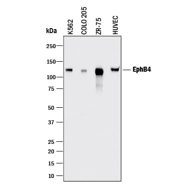
Detection of Human EphB4 by Western Blot. Western blot shows lysates of K562 human chronic myelogenous leukemia cell line, COLO 205 human colorectal adenocarcinoma cell line, ZR-75 human breast cancer cell line, and HUVEC human umbilical vein endothelial cells. PVDF membrane was probed with 2 µg/mL of Goat Anti-Human EphB4 Antigen Affinity-purified Polyclonal Antibody (Catalog # AF3038) followed by HRP-conjugated Anti-Goat IgG Secondary Antibody (Catalog # HAF017). A specific band was detected for EphB4 at approximately 120 kDa (as indicated). This experiment was conducted under reducing conditions and using Immunoblot Buffer Group 1.
 View Larger
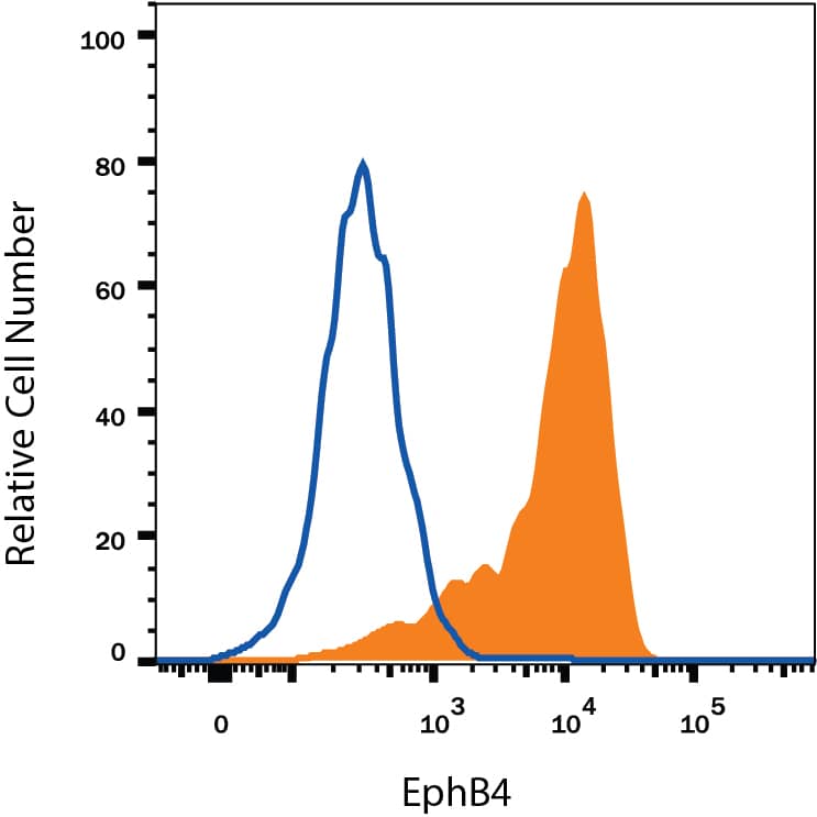
Detection of EphB4 in MCF‑7 Human Cell Line by Flow Cytometry. MCF-7 human breast cancer cell line was stained with Goat Anti-Human EphB4 Antigen Affinity-purified Polyclonal Antibody (Catalog # AF3038, filled histogram) or isotype control antibody (Catalog # AB-108-C, open histogram), followed by Phycoerythrin-conjugated Anti-Goat IgG Secondary Antibody (Catalog # F0107). View our protocol for Staining Membrane-associated Proteins.
 View Larger
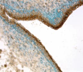
EphB4 in Human Kidney. EphB4 was detected in immersion fixed paraffin-embedded sections of human kidney using 15 µg/mL Goat Anti-Human EphB4 Antigen Affinity-purified Polyclonal Antibody (Catalog # AF3038) overnight at 4 °C. Tissue was stained with the Anti-Goat HRP-DAB Cell & Tissue Staining Kit (brown; Catalog # CTS008) and counterstained with hematoxylin (blue). View our protocol for Chromogenic IHC Staining of Paraffin-embedded Tissue Sections.
 View Larger
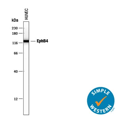
Detection of Human EphB4 by Simple WesternTM. Simple Western lane view shows lysates of HUVEC human umbilical vein endothelial cells, loaded at 0.2 mg/mL. A specific band was detected for EphB4 at approximately 127 kDa (as indicated) using 50 µg/mL of Goat Anti-Human EphB4 Antigen Affinity-purified Polyclonal Antibody (Catalog # AF3038) followed by 1:50 dilution of HRP-conjugated Anti-Goat IgG Secondary Antibody (Catalog # HAF109). This experiment was conducted under reducing conditions and using the 12-230 kDa separation system.
 View Larger
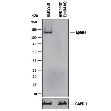
Western Blot Shows Human EphB4 Specificity by Using Knockout Cell Line. Western blot shows lysates of HEK293T human embryonic kidney parental cell line and EphB4 knockout HEK293T cell line (KO). PVDF membrane was probed with 2 µg/mL of Goat Anti-Human EphB4 Antigen Affinity-purified Polyclonal Antibody (Catalog # AF3038) followed by HRP-conjugated Anti-Goat IgG Secondary Antibody (Catalog # HAF017). A specific band was detected for EphB4 at approximately 140 kDa (as indicated) in the parental HEK293T cell line, but is not detectable in knockout HEK293T cell line. GAPDH (Catalog # AF5718) is shown as a loading control. This experiment was conducted under reducing conditions and using Immunoblot Buffer Group 1.
 View Larger
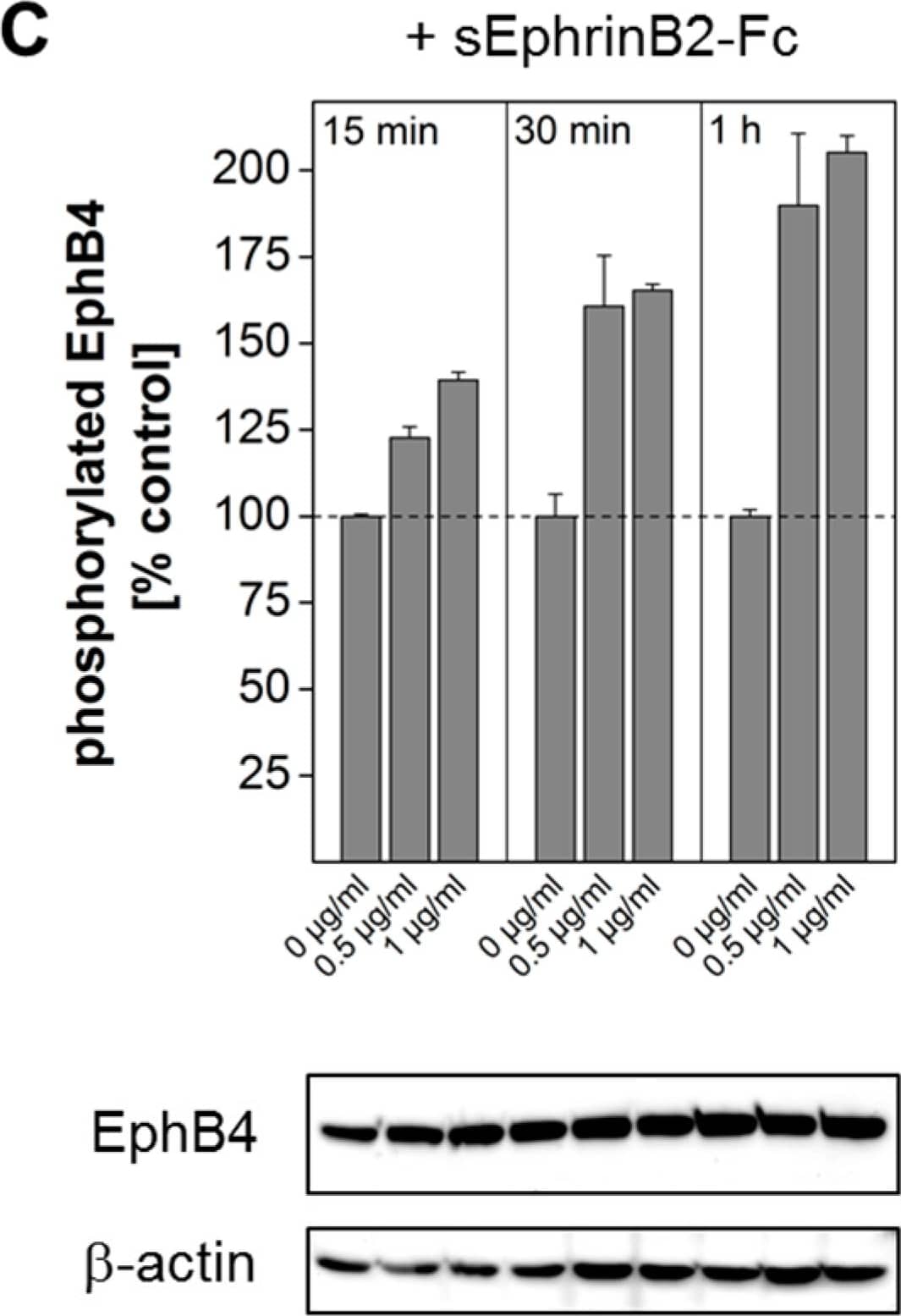
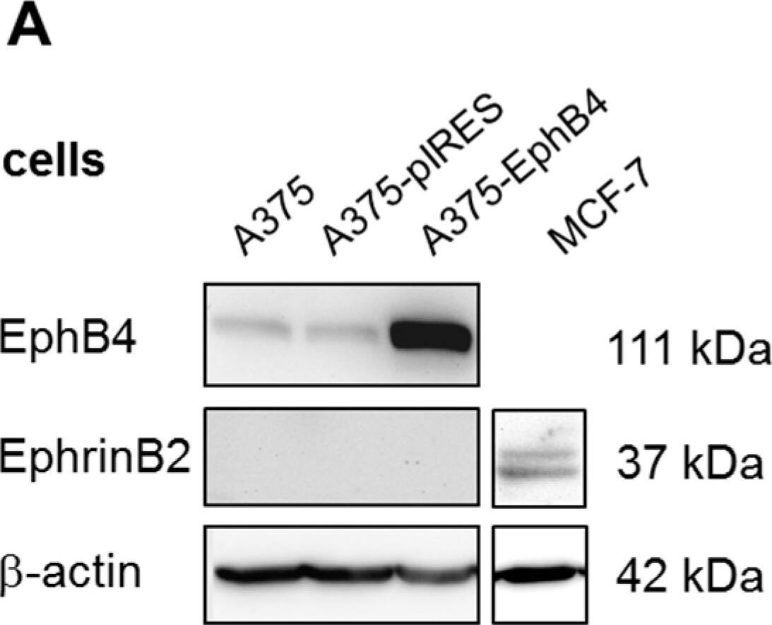
Detection of Mouse EphB4 by Western Blot EphB4 expression in transfected A375 melanoma cells and corresponding tumor xenografts. Western blot analysis of EphB4 and its ligand EphrinB2 in A375, A375-pIRES, and A375-EphB4 whole cell lysates (A) and tumor lysates (E). Anti-beta -actin served as loading control. (B) Relative mRNA expression of EphB4 and EphrinB2 in A375, A375-pIRES, and A375-EphB4 cells, analyzed by quantitative real-time RT-PCR was normalized to the constitutive expression level of beta -actin and to expression level in wild-type A375 melanoma cells using the delta delta CT method (2−∆∆ct) resulting in a value of 1 for A375 cells. Values represent mean ± SEM of at least three independent experiments each performed in triplicate (* p < 0.05, *** p < 0.001). (C) EphB4 phosphorylation was analyzed by pEphB4-ELISA in whole cell lysates of A375 EphB4 cells incubated with different concentrations of sEphrinB2-Fc (0; 0,5; 1 µg/mL) for 15, 30, and 60 min. Values represent mean ± SD from one of at least three independent experiments, each performed in duplicate. To rule out influence of sEphrinB2 on total EphB4 protein amount, EphB4 was analyzed in the same cell lysates by western blot analysis (figure shows one representative blot out of three independent experiments performed in duplicate) ranging from 92% to 112% of control as calculated after densitometric analysis. Anti beta -actin served as loading control. (D) Immunohistochemical detection of EphB4 in acetone-fixed cryosections of A375 pIRES and A375 EphB4 tumor xenografts using goat anti EphB4 antibody. Sections stained without the primary antibody served as negative control. Scale bar 50 µm. Image collected and cropped by CiteAb from the following publication (https://pubmed.ncbi.nlm.nih.gov/29462967), licensed under a CC-BY license. Not internally tested by R&D Systems.
Preparation and Storage
- 12 months from date of receipt, -20 to -70 °C as supplied.
- 1 month, 2 to 8 °C under sterile conditions after reconstitution.
- 6 months, -20 to -70 °C under sterile conditions after reconstitution.
Background: EphB4
EphB4, also known as Htk, Myk1, Tyro11, and Mdk2, is a member of the Eph receptor tyrosine kinase family and binds Ephrin-B2. The A and B class Eph proteins have a common structural organization (1-4). The human EphB4 cDNA encodes a 987 amino acid (aa) precursor that includes a 15 aa signal sequence, a 524 aa extracellular domain (ECD), a 21 aa transmembrane segment, and a 427 aa cytoplasmic domain (5). The ECD contains an N-terminal globular domain, a cysteine-rich domain, and two fibronectin type III domains. The cytoplasmic domain contains a juxtamembrane motif with two tyrosine residues which are the major autophosphorylation sites, a kinase domain, and a conserved sterile alpha motif (SAM) (5). Activation of kinase activity occurs after membrane-bound or clustered ligand recognition and binding. The ECD of human EphB4 shares 89% aa sequence identity with mouse EphB4 and 42-45% aa sequence identity with human EphB1, 2, and 3. EphB4 is expressed preferentially on venous endothelial cells (EC) and inhibits cell-cell adhesion, chemotaxis, and angiogenesis. Opposing effects are induced by signaling through Ephrin-B2 expressed on arterial EC: adhesion, endothelial cell migration, and vessel sprouting (6). EphB4 singaling contributes to new vascularization by guiding venous EC away from Ephrin-B2 expressing EC. Ephrin-B2 signaling induces arterial EC to migrate towards nascent EphB4 expressing vessels (6). The combination of forward signaling through EphB4 and reverse signaling through Ephrin-B2 promotes in vivo mammary tumor growth and
tumor-associated angiogenesis (7). EphB4 promotes the differentiation of megakaryocytic and erythroid progenitors but not granulocytic or monocytic progenitors (8, 9).
- Poliakov, A. et al. (2004) Dev. Cell 7:465.
- Surawska, H. et al. (2004) Cytokine Growth Factor Rev. 15:419.
- Pasquale, E.B. (2005) Nat. Rev. Mol. Cell Biol. 6:462.
- Davy, A. and P. Soriano (2005) Dev. Dyn. 232:1.
- Bennett, B.D. et al. (1994) J. Biol. Chem. 269:14211.
- Fuller, T. et al. (2003) J. Cell Sci. 116:2461.
- Noren, N.K. et al. (2004) Proc. Natl. Acad. Sci. USA 101:5583.
- Wang, Z. et al. (2002) Blood 99:2740.
- Inada, T. et al. (1997) Blood 89:2757.
Product Datasheets
Citations for Human EphB4 Antibody
R&D Systems personnel manually curate a database that contains references using R&D Systems products. The data collected includes not only links to publications in PubMed, but also provides information about sample types, species, and experimental conditions.
14
Citations: Showing 1 - 10
Filter your results:
Filter by:
-
EphrinB2-EphB4 signalling provides Rho-mediated homeostatic control of lymphatic endothelial cell junction integrity
Authors: Maike Frye, Simon Stritt, Henrik Ortsäter, Magda Hernandez Vasquez, Mika Kaakinen, Andres Vicente et al.
eLife
-
Evaluation of EphB4 as Target for Image-Guided Surgery of Breast Cancer
Authors: Cansu de Muijnck, Yoren van Gorkom, Maurice van Duijvenvoorde, Mina Eghtesadi, Geeske Dekker-Ensink, Shadhvi S. Bhairosingh et al.
Pharmaceuticals (Basel)
-
Janus-faced EPHB4-associated disorders: novel pathogenic variants and unreported intrafamilial overlapping phenotypes
Authors: Silvia Martin-Almedina, Kazim Ogmen, Ege Sackey, Dionysios Grigoriadis, Christina Karapouliou, Noeline Nadarajah et al.
Genetics in Medicine
-
Placenta Percreta Presents with Neoangiogenesis of Arteries with Von Willebrand Factor-Negative Endothelium
Authors: Alexander Schwickert, Wolfgang Henrich, Martin Vogel, Kerstin Melchior, Loreen Ehrlich, Matthias Ochs et al.
Reproductive Sciences
-
An EPHB4-RASA1 signaling complex inhibits shear stress-induced Ras-MAPK activation in lymphatic endothelial cells to promote the development of lymphatic vessel valves
Authors: Chen, D;Wiggins, D;Sevick, EM;Davis, MJ;King, PD;
bioRxiv : the preprint server for biology
Species: Human
Sample Types: Cell Lysates
Applications: Western Blot -
Hypoxia Triggers the Intravasation of Clustered Circulating Tumor Cells
Authors: C Donato, L Kunz, F Castro-Gin, A Paasinen-S, K Strittmatt, BM Szczerba, R Scherrer, N Di Maggio, W Heusermann, O Biehlmaier, C Beisel, M Vetter, C Rochlitz, WP Weber, A Banfi, T Schroeder, N Aceto
Cell Rep, 2020-09-08;32(10):108105.
Species: Human, Mouse
Sample Types: Cell Lysates
Applications: Western Blot -
Downregulation of proinflammatory cytokines in HTLV-1-infected T cells by Resveratrol
J Exp Clin Cancer Res, 2016-07-22;35(1):118.
Species: Human
Sample Types: Cell Lysates
Applications: Immunoprecipitation, Western Blot -
EPHB4 kinase-inactivating mutations cause autosomal dominant lymphatic-related hydrops fetalis
J Clin Invest, 2016-07-11;0(0):.
Species: Human
Sample Types: Cell Lysates
Applications: Immunoprecipitation, Western Blot -
Instruction of circulating endothelial progenitors in vitro towards specialized blood-brain barrier and arterial phenotypes.
Authors: Boyer-Di Ponio J, El-Ayoubi F, Glacial F, Ganeshamoorthy K, Driancourt C, Godet M, Perriere N, Guillevic O, Couraud P, Uzan G
PLoS ONE, 2014-01-02;9(1):e84179.
Species: Human
Sample Types: Whole Cells
Applications: ICC -
BCR-ABL-independent and RAS / MAPK pathway-dependent form of imatinib resistance in Ph-positive acute lymphoblastic leukemia cell line with activation of EphB4.
Authors: Suzuki M, Abe A, Imagama S, Nomura Y, Tanizaki R, Minami Y, Hayakawa F, Ito Y, Katsumi A, Yamamoto K, Emi N, Kiyoi H, Naoe T
Eur. J. Haematol., 2009-11-28;84(3):229-38.
Species: Human
Sample Types: Cell Lysates
Applications: Western Blot -
Soluble EphB4 inhibition of PDGF-induced RPE migration in vitro.
Authors: He S, Kumar SR, Zhou P, Krasnoperov V, Ryan SJ, Gill PS, Hinton DR
Invest. Ophthalmol. Vis. Sci., 2009-08-20;51(1):543-52.
Species: Human
Sample Types: Whole Tissue
Applications: IHC-Fr -
Activation of the receptor EphB4 by its specific ligand ephrin B2 in human osteoarthritic subchondral bone osteoblasts.
Authors: Kwan Tat S, Pelletier JP, Amiable N, Boileau C, Lajeunesse D, Duval N, Martel-Pelletier J
Arthritis Rheum., 2008-12-01;58(12):3820-30.
Species: Human
Sample Types: Whole Tissue
Applications: IHC -
Evaluation of EphA2 and EphB4 as Targets for Image-Guided Colorectal Cancer Surgery
Authors: Marieke A. Stammes, Hendrica A. J. M. Prevoo, Meyke C. Ter Horst, Stéphanie A. Groot, Cornelis J. H. Van de Velde, Alan B. Chan et al.
International Journal of Molecular Sciences
-
Overexpression of Receptor Tyrosine Kinase EphB4 Triggers Tumor Growth and Hypoxia in A375 Melanoma Xenografts: Insights from Multitracer Small Animal Imaging Experiments.
Authors: Neuber C, Belter B, Meister S, Hofheinz F.
Molecules.
FAQs
No product specific FAQs exist for this product, however you may
View all Antibody FAQsReviews for Human EphB4 Antibody
Average Rating: 5 (Based on 1 Review)
Have you used Human EphB4 Antibody?
Submit a review and receive an Amazon gift card.
$25/€18/£15/$25CAN/¥75 Yuan/¥2500 Yen for a review with an image
$10/€7/£6/$10 CAD/¥70 Yuan/¥1110 Yen for a review without an image
Filter by:

