Human Galectin-7 Antibody Summary
Ser2-Phe136
Accession # NP_002298
Applications
Human Galectin-7 Sandwich Immunoassay
Please Note: Optimal dilutions should be determined by each laboratory for each application. General Protocols are available in the Technical Information section on our website.
Scientific Data
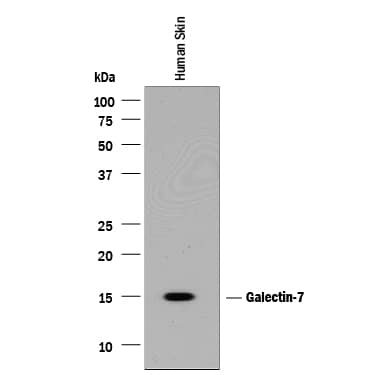 View Larger
Detection of Human Galectin‑7 by Western Blot. Western blot shows lysates of human skin tissue. PVDF membrane was probed with 1 µg/mL of Goat Anti-Human Galectin-7 Antigen Affinity-purified Polyclonal Antibody (Catalog # AF1339) followed by HRP-conjugated Anti-Goat IgG Secondary Antibody (Catalog # HAF017). A specific band was detected for Galectin-7 at approximately 15 kDa (as indicated). This experiment was conducted under reducing conditions and using Immunoblot Buffer Group 1.
 View Larger
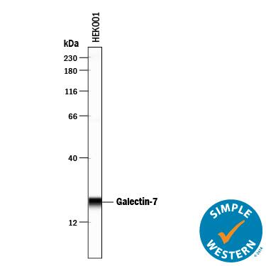
Detection of Human Galectin‑7 by Simple WesternTM. Simple Western lane view shows lysates of HEK001 human epidermal keratinocyte cell line, loaded at 0.2 mg/mL. A specific band was detected for Galectin-7 at approximately 21 kDa (as indicated) using 10 µg/mL of Goat Anti-Human Galectin-7 Antigen Affinity-purified Polyclonal Antibody (Catalog # AF1339) followed by 1:50 dilution of HRP-conjugated Anti-Goat IgG Secondary Antibody (Catalog # HAF109). This experiment was conducted under reducing conditions and using the 12-230 kDa separation system.
Reconstitution Calculator
Preparation and Storage
- 12 months from date of receipt, -20 to -70 °C as supplied.
- 1 month, 2 to 8 °C under sterile conditions after reconstitution.
- 6 months, -20 to -70 °C under sterile conditions after reconstitution.
Background: Galectin-7
The galectins constitute a large family of carbohydrate-binding proteins with specificity for N-acetyl-lactosamine-containing glycoproteins. At least 14 mammalian galectins, which share structural similarities in their carbohydrate recognition domains (CRD), have been identified. The galectins have been classified into the prototype galectins (-1, -2, -5, -7, -10, -11, -13, -14), which contain one CRD and exist either as a monomer or a noncovalent homodimer; the chimera galectins (Galectin-3) containing one CRD linked to a nonlectin domain; and the tandem-repeat galectins (-4, -6, -8, -9, -12) consisting of two CRDs joined by a linker peptide. Galectins lack a classical signal peptide and can be localized to the cytosolic compartments where they have intracellular functions. However, via one or more as yet unidentified non-classical secretory pathways, galectins can also be secreted to function extracellularly. Individual members of the galectin family have different tissue distribution profiles and exhibit subtle differences in their carbohydrate-binding specificities. Each family member may preferentially bind to a unique subset of cell-surface glycoproteins (1-4). Human Galectin-7 is a prototype monomeric galectin. It is specifically expressed in stratified epithelia, notably in epidermis, but is barely detectable in epidermal tumors and significantly down regulated or absent from squamous carconima cell lines. The Galectin-7 gene is induced by tumor suppressor protein p53 transcriptional activity following genotoxic events. A pro-apoptotic protein, Galectin-7 functions intracellularly upstream of JNK activation and cytochrome-c release. This protein has been shown to increase the susceptibility of keratinocytes to UVB induced apoptosis, an essential processss in the maintenance of epidermal homeostasis. Cell lines transfected with the Galectin-7 gene localized the protein in the nucleus and intracellularly. Human and mouse Galectin-7 share 79% amino acid homology (4-6).
- Rabinovich, A. et al. (2002) TRENDS in Immunol. 23:313.
- Rabinovich, A. et al. (2002) J. Leukocyte Biology 71:741.
- Hughes, R.C. (2002) Biochimie 83:667.
- R&D Systems Cytokine Bulletin; Summer 2002.
- Bernerd, F. et al. (1999) Proc. Natl. Acad. Sci. USA 96:11329.
- Kuwabara, I. et al. (2002) J. Biol. Chem. 277:3487.
Product Datasheets
Citations for Human Galectin-7 Antibody
R&D Systems personnel manually curate a database that contains references using R&D Systems products. The data collected includes not only links to publications in PubMed, but also provides information about sample types, species, and experimental conditions.
5
Citations: Showing 1 - 5
Filter your results:
Filter by:
-
Astrocytic response mediated by the CLU risk allele inhibits OPC proliferation and myelination in a human iPSC model
Authors: Zhenqing Liu, Jianfei Chao, Cheng Wang, Guihua Sun, Daniel Roeth, Wei Liu et al.
Cell Reports
-
Tissue and plasma levels of galectins in patients with high grade serous ovarian carcinoma as new predictive biomarkers
Authors: M Labrie, LOF De Araujo, L Communal, AM Mes-Masson, Y St-Pierre
Sci Rep, 2017-10-16;7(1):13244.
Species: Human
Sample Types: Whole Tissue
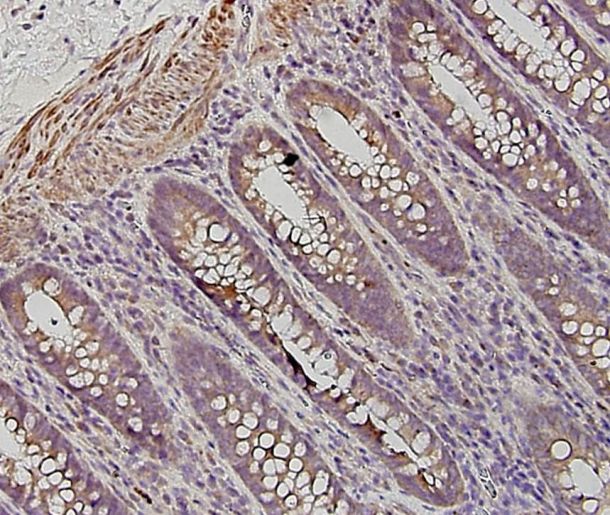
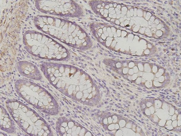
Applications: IHC-P -
A Mutation in the Carbohydrate Recognition Domain Drives a Phenotypic Switch in the Role of Galectin-7 in Prostate Cancer.
Authors: Labrie M, Vladoiu M, Leclerc B, Grosset A, Gaboury L, Stagg J, St-Pierre Y
PLoS ONE, 2015-07-13;10(7):e0131307.
Species: Human
Sample Types: Whole Tissue
Applications: IHC-P -
The CCAAT/enhancer-binding protein beta-2 isoform (CEBPbeta-2) upregulates galectin-7 expression in human breast cancer cells.
Authors: Campion C, Labrie M, Grosset A, St-Pierre Y
PLoS ONE, 2014-05-02;9(5):e95087.
Species: Human
Sample Types: Whole Cells
Applications: ICC -
Galectin-7 acts as an adhesion molecule during implantation and increased expression is associated with miscarriage.
Authors: Menkhorst E, Gamage T, Cuman C, Kaitu'u-Lino T, Tong S, Dimitriadis E
Placenta, 2014-01-24;35(3):195-201.
Species: Human
Sample Types: Whole Tissue
Applications: IHC
FAQs
No product specific FAQs exist for this product, however you may
View all Antibody FAQsReviews for Human Galectin-7 Antibody
Average Rating: 4.3 (Based on 4 Reviews)
Have you used Human Galectin-7 Antibody?
Submit a review and receive an Amazon gift card.
$25/€18/£15/$25CAN/¥75 Yuan/¥2500 Yen for a review with an image
$10/€7/£6/$10 CAD/¥70 Yuan/¥1110 Yen for a review without an image
Filter by:
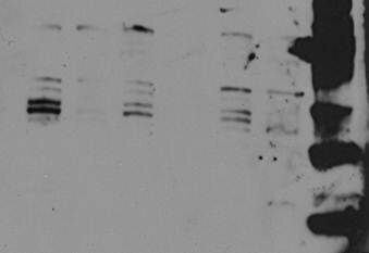
First run with 1:1000 for difference cancer cell lysates gave many bands, which could be isoforms detected.
Specificity: Reasonably specific
Sensitivity: Reasonably sensitive
Buffer: BSA
Dilution: 1:1000
At higher dilutions, only the main form was visible.
Specificity: Specific
Sensitivity: Sensitive
Buffer: Loading buffer
Dilution: 1:2000
At 1:500 the antibody is very efficient in detecting galectin-7.
Specificity: Specific
Sensitivity: Sensitive
Buffer: PBS
Dilution: 1:500
