Human GLI-3 Antibody Summary
Met1-Glu479
Accession # P10071
Applications
Please Note: Optimal dilutions should be determined by each laboratory for each application. General Protocols are available in the Technical Information section on our website.
Scientific Data
 View Larger
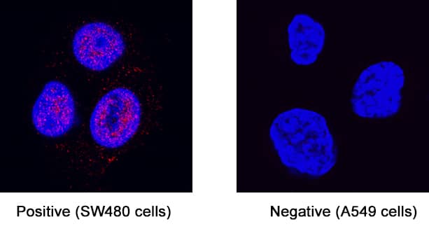
Detection of GLI-3 in Human SW480 cell line. GLI‑3 was detected in immersion fixed SW480 fibroblast carcinoma cell line (positive staining) and A549 human lung carcinoma cell line (negative staining) using Rabbit Anti-Human GLI‑3 Monoclonal Antibody (Catalog # MAB3690) at 3 µg/mL for 3 hours at room temperature. Cells were stained using the NorthernLights™ 557-conjugated Anti-Rabbit IgG Secondary Antibody (red; Catalog # NL004) and counterstained with DAPI (blue). Specific staining was localized to cell nuclei. View our protocol for Fluorescent ICC Staining of Cells on Coverslips.
Reconstitution Calculator
Preparation and Storage
- 12 months from date of receipt, -20 to -70 °C as supplied.
- 1 month, 2 to 8 °C under sterile conditions after reconstitution.
- 6 months, -20 to -70 °C under sterile conditions after reconstitution.
Background: GLI-3
GLI3 is a zinc finger protein. The GLI family of proteins are transcription factors and are mediators of Sonic hedgehog signaling. GLI3 is known to be a transcriptional repressor but also may have a positive transcription function. Mutations in the GLI3 gene are associated with many polydactyly diseases.
Product Datasheets
FAQs
No product specific FAQs exist for this product, however you may
View all Antibody FAQsReviews for Human GLI-3 Antibody
There are currently no reviews for this product. Be the first to review Human GLI-3 Antibody and earn rewards!
Have you used Human GLI-3 Antibody?
Submit a review and receive an Amazon gift card.
$25/€18/£15/$25CAN/¥75 Yuan/¥2500 Yen for a review with an image
$10/€7/£6/$10 CAD/¥70 Yuan/¥1110 Yen for a review without an image


