Human IL-18/IL-1F4 Propeptide Antibody Summary
Applications
Please Note: Optimal dilutions should be determined by each laboratory for each application. General Protocols are available in the Technical Information section on our website.
Scientific Data
 View Larger
Detection of Human IL‑18/IL‑1F4 by Western Blot. Western blot shows lysates of PC-3 human prostate cancer cell line, MCF 10A human breast epithelial cell line, A431 human epithelial carcinoma cell line, and HEK293T human embryonic kidney cell line (negative control cell line). PVDF membrane was probed with 0.5 µg/mL of Goat Anti-Human IL-18/IL-1F4 Propeptide Antigen Affinity-purified Polyclonal Antibody (Catalog # AF646) followed by HRP-conjugated Anti-Goat IgG Secondary Antibody (Catalog # HAF017). A specific band was detected for IL-18/IL-1F4 at approximately 22 kDa (as indicated). GAPDH (Catalog # AF5718) is shown as a loading control. This experiment was conducted under reducing conditions and using Immunoblot Buffer Group 1.
 View Larger
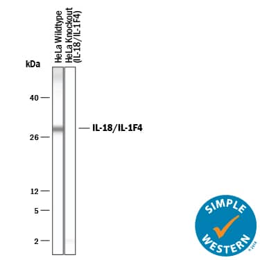
Detection of Human IL‑18/IL‑1F4 by Simple WesternTM. Simple Western lane view shows lysates of HeLa human cervical epithelial carcinoma parental cell line and IL-18/IL-1F4 knockout HeLa cell line, loaded at 0.2 mg/mL. A specific band was detected for IL-18/IL-1F4 at approximately 29 kDa (as indicated) in the HeLa parental cell line using 50 µg/mL of Goat Anti-Human IL-18/IL-1F4 Propeptide Antigen Affinity-purified Polyclonal Antibody (Catalog # AF646) followed by 1:50 dilution of HRP-conjugated Anti-Goat IgG Secondary Antibody (Catalog # HAF109). This experiment was conducted under reducing conditions and using the 12-230 kDa separation system.
 View Larger
Western Blot Shows Human IL‑18/IL‑1F4 Specificity by Using Knockout Cell Line. Western blot shows lysates of HeLa human cervical epithelial carcinoma parental cell line and IL-18/IL-1F4 knockout HeLa cell line (KO). PVDF membrane was probed with 0.5 µg/mL of Goat Anti-Human IL-18/IL-1F4 Propeptide Antigen Affinity-purified Polyclonal Antibody (Catalog # AF646) followed by HRP-conjugated Anti-Goat IgG Secondary Antibody (Catalog # HAF017). A specific band was detected for IL-18/IL-1F4 at approximately 22 kDa (as indicated) in the parental HeLac ell line, but is not detectable in knockout HeLa cell line. GAPDH (Catalog # AF5718) is shown as a loading control. This experiment was conducted under reducing conditions and using Immunoblot Buffer Group 1.
 View Larger
Detection of Rhesus Macaque IL-18/IL-1F4 by Immunohistochemistry CD8+ T- and CD20+ B-lymphocytes are not localized within inflammatory foci.Immunohistochemistry using anti-CD20 identified a few B-lymphocytes in the inflammatory foci (arrows) (A). In contrast, no CD8+ T-lymphocytes are localized using anti-CD8 antibody (B) (400X). Immunohistrochemistry using anti-pro-IL-18 (C) or anti-CXCL9 (D) reveals the expression of these molecules on mononuclear cells within inflammatory foci (200X). Image collected and cropped by CiteAb from the following open publication (https://dx.plos.org/10.1371/journal.pone.0014429), licensed under a CC-BY license. Not internally tested by R&D Systems.
Reconstitution Calculator
Preparation and Storage
- 12 months from date of receipt, -20 to -70 °C as supplied.
- 1 month, 2 to 8 °C under sterile conditions after reconstitution.
- 6 months, -20 to -70 °C under sterile conditions after reconstitution.
Background: IL-18/IL-1F4
Pro-IL-18 (pro-Interleukin 18; also pro-IGIF and pro-IL-1 gamma ) is a 24 kDa member of the IL-1 family of molecules. It is widely expressed, being produced by keratinocytes, intestinal epithelium, T cells, macrophages and osteoblasts. Human Pro-IL-18 is 193 amino acids (aa) in length. Although mature IL-18 induces IFN-gamma secretion by NK and T cells, Pro-IL-18 appears to have little intrinsic activity. Generally, active IL-18 is considered to arise from caspase-1 cleavage of Pro-IL-18 between Asp36-Tyr37. This generates an 18 kDa mature C-terminal fragment, and a 4 kDa (predicted) N-terminal prosegment that runs at 6 kDa in SDS-PAGE. Other proteases are known to process Pro-IL-18. Caspase-3 cleavage after Asp68 generates an inactive 14 kDa mature segment, Merpin beta -subunit cleavage after Asn52 generates a marginally active 17 kDa mature segment, while parasite Cys protease cleavage after Val47 generates an inactive 17 kDa mature molecule. One splice variant shows a deletion of aa 27-30. Over aa 2-36, human Pro-IL-18 shares 63% aa identity with mouse Pro-IL-18.
Product Datasheets
Citations for Human IL-18/IL-1F4 Propeptide Antibody
R&D Systems personnel manually curate a database that contains references using R&D Systems products. The data collected includes not only links to publications in PubMed, but also provides information about sample types, species, and experimental conditions.
3
Citations: Showing 1 - 3
Filter your results:
Filter by:
-
Myocarditis in CD8-depleted SIV-infected rhesus macaques after short-term dual therapy with nucleoside and nucleotide reverse transcriptase inhibitors.
Authors: Annamalai L, Westmoreland SV, Domingues HG
PLoS ONE, 2010-12-23;5(12):e14429.
Species: Primate - Macaca mulatta (Rhesus Macaque)
Sample Types: Whole Tissue
Applications: IHC-P -
Differentiation of human SH-SY5Y neuroblastoma cells by all-trans retinoic acid activates the interleukin-18 system.
Authors: Sallmon H, Hoene V, Weber SC, Dame C
J. Interferon Cytokine Res., 2010-02-01;30(2):55-8.
Species: Human
Sample Types: Whole Cells
Applications: ICC -
Interleukin-18 predicts atherosclerosis progression in SIV-infected and uninfected rhesus monkeys (Macaca mulatta) on a high-fat/high-cholesterol diet.
Authors: Yearley JH, Xia D, Pearson CB, Carville A, Shannon RP, Mansfield KG
Lab. Invest., 2009-04-20;89(6):657-67.
Species: Primate - Macaca mulatta (Rhesus Macaque)
Sample Types: Whole Tissue
Applications: IHC-P
FAQs
No product specific FAQs exist for this product, however you may
View all Antibody FAQsReviews for Human IL-18/IL-1F4 Propeptide Antibody
There are currently no reviews for this product. Be the first to review Human IL-18/IL-1F4 Propeptide Antibody and earn rewards!
Have you used Human IL-18/IL-1F4 Propeptide Antibody?
Submit a review and receive an Amazon gift card.
$25/€18/£15/$25CAN/¥75 Yuan/¥2500 Yen for a review with an image
$10/€7/£6/$10 CAD/¥70 Yuan/¥1110 Yen for a review without an image


