Human Indoleamine 2,3-dioxygenase/IDO PE-conjugated Antibody
Human Indoleamine 2,3-dioxygenase/IDO PE-conjugated Antibody Summary
Ala2-Gly403
Accession # P14902
Customers also Viewed
Applications
Please Note: Optimal dilutions should be determined by each laboratory for each application. General Protocols are available in the Technical Information section on our website.
Scientific Data
 View Larger
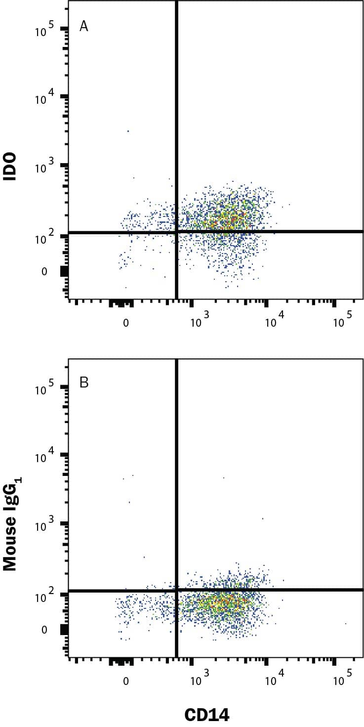
Detection of Indoleamine 2,3‑dioxygenase/IDO in Human Monocytes by Flow Cytometry. Human Monocytes were selected from PBMC using MagCellect Human CD14+ Cell Isolation Kit (MAGH105) and cultured overnight with Recombinant Human MCSF (50 ng/mL; 216-MC), Recombinant Human IFN gamma (50 ng/mL; 285-IF) and 50 ng/mL LPS and stained with (A) Mouse Anti-Human Indoleamine 2,3-dioxygenase/IDO PE-conjugated Monoclonal Antibody (Catalog # IC6030P) or (B) Mouse IgG1 isotype control antibody (IC002P) and Mouse Anti-Human CD14 APC-conjugated Monoclonal Antibody (FAB3832A). To facilitate intracellular staining, cells were fixed and permeabilized with FlowX FoxP3/Transcription Factor Fixation & Perm Kit (FC012). Staining was performed using our Staining Intracellular Molecules protocol.
 View Larger
Detection of Indoleamine 2,3‑dioxygenase/IDO in Human MDSCs by Flow Cytometry. Human myeloid-derived suppressor cells (MDSCs) treated with 10 ng/mL Recombinant Human IL-6 (Catalog # 206-IL) and 10 ng/mL Recombinant Human GM-CSF (Catalog # 215-GM) for 7 days were stained with Mouse Anti-Human Siglec-3/CD33 APC-conjugated Monoclonal Antibody (Catalog # FAB1137A) and either (A) Mouse Anti-Human Indoleamine 2,3-dioxygenase/IDO PE-conjugated Monoclonal Antibody (Catalog # IC6030P) or (B) Mouse IgG1Phycoerythrin Isotype Control (Catalog # IC002P). To facilitate intracellular staining, cells were fixed with Flow Cytometry Fixation Buffer (Catalog # FC004) and permeabilized with Flow Cytometry Permeabilization/Wash Buffer I (Catalog # FC005). View our protocol for Staining Intracellular Molecules.
 View Larger
Detection of Human Indoleamine 2,3-dioxygenase/IDO by Flow Cytometry SLE sera increase IDO expression in a type I IFN-dependent manner.Endothelial cells (WISH cells) were incubated with A) recombinant IFN-alpha with or without pre-incubation with an IFN alpha/beta receptor blocking antibody (IFNAR ab) and IDO expression analyzed in permeabilized cells by flow cytometry. B) WISH cells were incubated with serum from SLE patients or healthy individuals with or without pre-incubation with an IFNAR ab. The results are presented as percentage increased IDO expression in median fluorescence intensity (MFI) as compared to non-stimulated cells. C) Patients with ongoing type I IFN activity (SLE IFN+) had increased IDO activity as compared to patients with no type I IFN activity (SLE IFN-) and healthy volunteers (HV). D) Patients with ongoing type I IFN activity had decreased serotonin levels as compared to patients with no type I IFN activity and healthy volunteers. Image collected and cropped by CiteAb from the following publication (https://dx.plos.org/10.1371/journal.pone.0125109), licensed under a CC-BY license. Not internally tested by R&D Systems.
Preparation and Storage
- 12 months from date of receipt, 2 to 8 °C as supplied.
Background: Indoleamine 2,3-dioxygenase/IDO
Indoleamine 2,3-dioxygenase (also known as IDO) is a heme-containing intracellular dioxygenase catalyzing the degradation of the essential amino acid L-tryptophan to N‑formyl‑kynurenine (1). This degradation is the first and rate-limiting step of the L-kynurenine pathway (2). IDO is widely expressed in dendritic cells, macrophages, microglia, eosinophils, fibroblasts, endothelial cells, and most tumor cells. In immune cells, its expression is mainly induced by cytokines such as IFN-gamma, IFN-alpha, IFN‑ beta, and IL-10. IDO has an antimicrobial function due to its decreasing the availability of the essential amino acid tryptophan in inflammatory environments (3). Recent studies have demonstrated that IDO induces immunosuppression during infection, pregnancy, transplantation, autoimmunity, and neoplasia (3-5).
- Lewis-Ballester, A. et al. (2009) Proc. Natl. Acad. Sci. USA. 106:17371.
- Costantino, G. (2009) Expert Opin. Ther. Targets 13:247.
- Xu, H. et al. (2008) Immunol. Lett. 121:1.
- Lob, S. et al. (2009) Nat. Rev. Cancer 9:445.
- Curti, A. et al. (2009) Blood 113:2394.
Product Datasheets
Citations for Human Indoleamine 2,3-dioxygenase/IDO PE-conjugated Antibody
R&D Systems personnel manually curate a database that contains references using R&D Systems products. The data collected includes not only links to publications in PubMed, but also provides information about sample types, species, and experimental conditions.
6
Citations: Showing 1 - 6
Filter your results:
Filter by:
-
High M-MDSC Percentage as a Negative Prognostic Factor in Chronic Lymphocytic Leukaemia
Authors: M Zarobkiewi, W Kowalska, S Chocholska, W Tomczak, A Szyma?ska, I Morawska, A Wojciechow, A Bojarska-J
Cancers (Basel), 2020-09-14;12(9):.
-
Preconditioned Chorionic Villus Mesenchymal Stem/Stromal Cells (CVMSCs) Minimize the Invasive Phenotypes of Breast Cancer Cell Line MDA231 In Vitro
Authors: Subayyil, AA;Basmaeil, YS;Kulayb, HB;Alrodayyan, M;Alhaber, LAA;Almanaa, TN;Khatlani, T;
International journal of molecular sciences
Species: Human
Sample Types: Whole Cells
Applications: Flow Cytometry -
Mycobacterium leprae induces a tolerogenic profile in monocyte-derived dendritic cells via TLR2 induction of IDO
Authors: JAP Oliveira, M Gandini, JS Sales, SK Fujimori, MGM Barbosa, VS Frutuoso, MO Moraes, EN Sarno, MCV Pessolani, RO Pinheiro
J Leukoc Biol, 2020-10-11;0(0):.
Species: Human
Sample Types: Whole Cells
Applications: Flow Cytometry -
Mesenchymal Stromal Cells Are More Immunosuppressive In Vitro If They Are Derived from Endometriotic Lesions than from Eutopic Endometrium
Authors: F Abomaray, S Gidlöf, C Götherströ
Stem Cells Int, 2017-11-05;2017(0):3215962.
Species: Human
Sample Types: Whole Cells
Applications: Flow Cytometry -
Indoleamine 2, 3-dioxygenase (IDO) increases during renal fibrogenesis and its inhibition potentiates TGF-? 1-induced epithelial to mesenchymal transition
Authors: LHG Matheus, GM Simão, TA Amaral, RBO Brito, CS Malta, YST Matos, AC Santana, GGC Rodrigues, MC Albejante, EE Bach, MA Dalboni, CP Camacho, H Dellê
BMC Nephrol, 2017-09-06;18(1):287.
Species: Canine
Sample Types: Whole Cells
Applications: ICC -
Type I interferon-mediated skewing of the serotonin synthesis is associated with severe disease in systemic lupus erythematosus.
Authors: Lood C, Tyden H, Gullstrand B, Klint C, Wenglen C, Nielsen C, Heegaard N, Jonsen A, Kahn R, Bengtsson A
PLoS ONE, 2015-04-21;10(4):e0125109.
Species: Human
Sample Types: Whole Cells
Applications: Flow Cytometry
FAQs
No product specific FAQs exist for this product, however you may
View all Antibody FAQsIsotype Controls
Staining Reagents
Reviews for Human Indoleamine 2,3-dioxygenase/IDO PE-conjugated Antibody
There are currently no reviews for this product. Be the first to review Human Indoleamine 2,3-dioxygenase/IDO PE-conjugated Antibody and earn rewards!
Have you used Human Indoleamine 2,3-dioxygenase/IDO PE-conjugated Antibody?
Submit a review and receive an Amazon gift card.
$25/€18/£15/$25CAN/¥75 Yuan/¥2500 Yen for a review with an image
$10/€7/£6/$10 CAD/¥70 Yuan/¥1110 Yen for a review without an image


