Human KAP1 Antibody Summary
Lys266-Ser624
Accession # Q13263
Customers also Viewed
Applications
Please Note: Optimal dilutions should be determined by each laboratory for each application. General Protocols are available in the Technical Information section on our website.
Scientific Data
 View Larger
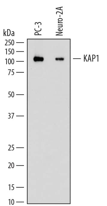
Detection of Human and Mouse KAP1 by Western Blot. Western blot shows lysates of PC-3 human prostate cancer cell line and Neuro-2A mouse neuroblastoma cell line. PVDF membrane was probed with 0.2 µg/mL of Mouse Anti-Human KAP1 Monoclonal Antibody (Catalog # MAB7785) followed by HRP-conjugated Anti-Mouse IgG Secondary Antibody (HAF018). A specific band was detected for KAP1 at approximately 105 kDa (as indicated). This experiment was conducted under reducing conditions and using Immunoblot Buffer Group 1.
 View Larger
Detection of Human KAP1 by Simple WesternTM. Simple Western lane view shows lysates of PC‑3 human prostate cancer cell line, loaded at 0.5 mg/mL. A specific band was detected for KAP1 at approximately 117 kDa (as indicated) using 2 µg/mL of Mouse Anti-Human KAP1 Monoclonal Antibody (Catalog # MAB7785). This experiment was conducted under reducing conditions and using the 12-230 kDa separation system.
 View Larger
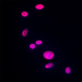
KAP1 in Neuro‑2A Mouse Cell Line. KAP1 was detected in immersion fixed Neuro-2A mouse neuroblastoma cell line using Mouse Anti-Human KAP1 Monoclonal Antibody (Catalog # MAB7785) at 8 µg/mL for 3 hours at room temperature. Cells were stained using the NorthernLights™ 557-conjugated Anti-Mouse IgG Secondary Antibody (red; NL007) and counterstained with DAPI (blue). Specific staining was localized to nuclei. View our protocol for Fluorescent ICC Staining of Cells on Coverslips.
 View Larger
KAP1 in Human Liver Cancer Tissue. KAP1 was detected in immersion fixed paraffin-embedded sections of human liver cancer tissue using Mouse Anti-Human KAP1 Monoclonal Antibody (Catalog # MAB7785) at 0.5 µg/mL for 1 hour at room temperature followed by incubation with the Anti-Mouse IgG VisUCyte™ HRP Polymer Antibody (VC001). Tissue was stained using DAB (brown) and counterstained with hematoxylin (blue). Specific staining was localized to nuclei. View our protocol for IHC Staining with VisUCyte HRP Polymer Detection Reagents.
 View Larger
Detection of KAP1 in PC‑3 human prostate cancer cells KAP1 was detected in immersion fixed PC‑3 human prostate cancer cells using Mouse Anti-Human KAP1 Monoclonal Antibody (Catalog # MAB7785) at 8 µg/mL for 3 hours at room temperature. Cells were stained using the NorthernLights™ 557-conjugated Anti-Mouse IgG Secondary Antibody (red; Catalog # NL007) and counterstained with DAPI (blue). Specific staining was localized to Nuclear. View our protocol for Fluorescent ICC Staining of Cells on Coverslips.
Preparation and Storage
- 12 months from date of receipt, -20 to -70 °C as supplied.
- 1 month, 2 to 8 °C under sterile conditions after reconstitution.
- 6 months, -20 to -70 °C under sterile conditions after reconstitution.
Background: KAP1
KAP1 (KRAB-interacting protein 1; gene name TRIM28), also called TIF1b (transcription intermediary factor 1b) is an approximately 100 kDa nuclear corepressor that is a member of the TRIM (tripartite motif-containing) family. It is reported to bind both IRF7 and the SUMO enzyme and cause increased SUMOylation of IRF7. The 834 amino acid (aa) mature protein contains four zinc finger regions and a leucine zipper domain. A 752 aa human splice variant lacking aa 114-195 has been reported. Within the region used as an immunogen, human KAP1 shares 94% aa sequence identity with mouse and rat KAP1.
Product Datasheets
FAQs
No product specific FAQs exist for this product, however you may
View all Antibody FAQsReviews for Human KAP1 Antibody
Average Rating: 5 (Based on 1 Review)
Have you used Human KAP1 Antibody?
Submit a review and receive an Amazon gift card.
$25/€18/£15/$25CAN/¥75 Yuan/¥2500 Yen for a review with an image
$10/€7/£6/$10 CAD/¥70 Yuan/¥1110 Yen for a review without an image
Filter by:


