Human LAMP-1/CD107a Antibody Summary
Ala28-Asn380
Accession # P11279
Applications
Please Note: Optimal dilutions should be determined by each laboratory for each application. General Protocols are available in the Technical Information section on our website.
Scientific Data
 View Larger
LAMP1/CD107a in Human Kidney. LAMP1/CD107a was detected in immersion fixed paraffin-embedded sections of human kidney using Mouse Anti-Human LAMP1/CD107a Monoclonal Antibody (Catalog # MAB4800) at 15 µg/mL overnight at 4 °C. Before incubation with the primary antibody, tissue was subjected to heat-induced epitope retrieval using Antigen Retrieval Reagent-Basic (Catalog # CTS013). Tissue was stained using the Anti-Mouse HRP-DAB Cell & Tissue Staining Kit (brown; Catalog # CTS002) and counterstained with hematoxylin (blue). Specific staining was localized to lysosomes in epithelial cells. View our protocol for Chromogenic IHC Staining of Paraffin-embedded Tissue Sections.
 View Larger
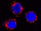
LAMP‑1/CD107a in THP‑1 Human Cell Line. LAMP-1/CD107a was detected in immersion fixed THP-1 human acute monocytic leukemia cell line using Mouse Anti-Human LAMP-1/CD107a Monoclonal Antibody (Catalog # MAB4800) at 25 µg/mL for 3 hours at room temperature. Cells were stained using the NorthernLights™ 557-conjugated Anti-Mouse IgG Secondary Antibody (red; Catalog # NL007) and counterstained with DAPI(blue). Specific staining was localized to cytoplasmic. View our protocol for Fluorescent ICC Staining of Cells on Coverslips.
 View Larger
Detection of Human LAMP-1/CD107a by Western Blot Increased SNX9 expression and co-localization with podocin are detectable in the cytoplasm of ADR-treated WT podocytes, whereas SNX9 KD podocytes exhibit little cytoplasmic expression of podocin.(a) Fluorescent micrographs of cultured human podocytes stained with SNX9 (green) and podocin (red) before and after ADR treatment (merged areas are in yellow). DAPI (blue) was used to indicate nuclei. Boxes indicate higher magnification areas presented in the lower panels. (b) Western blot analyses of the fractions from control podocytes or podocytes treated with ADR separated on linear OptiPrep gradients (5–25%). Distributions of SNX9 and podocin, as well as marker proteins of plasma membrane (caveolin), endosome/lysosome (LAMP1), mitochondria ( beta subunit of F1F0-ATPase), and endoplasmic reticulum (calnexin), were examined by western blot analysis. (c) Cultured human podocytes were transfected with nonfunctional control siRNA (upper panel) or SNX9 siRNA (middle and lower panels). Transfected cells, as decided by GFP expression, are indicated by arrowheads. Upper panel: Fluorescent micrographs of control siRNA-transfected podocyte stained with SNX9 (red). Middle panel: Fluorescent micrographs of SNX9 siRNA-transfected podocyte with ADR treatment stained with SNX9 (red). Lower panel: Fluorescent micrographs of SNX9 siRNA-transfected podocyte with ADR treatment stained with podocin (red). DAPI (blue) was used to indicate nuclei. Boxes indicate higher-magnification areas presented on the right. Image collected and cropped by CiteAb from the following publication (https://pubmed.ncbi.nlm.nih.gov/28266622), licensed under a CC-BY license. Not internally tested by R&D Systems.
Reconstitution Calculator
Preparation and Storage
- 12 months from date of receipt, -20 to -70 °C as supplied.
- 1 month, 2 to 8 °C under sterile conditions after reconstitution.
- 6 months, -20 to -70 °C under sterile conditions after reconstitution.
Background: LAMP-1/CD107a
Lysosome-associated membrane protein-1 (LAMP1), also known as CD107a, is a 100‑130 kDa member of the LAMP family of glycoproteins. It is expressed in lysosomal and plasma membranes of macrophages, NK and T-cells, and with LAMP2, is essential for the formation of phagolysosomes. On the cell surface, it also presents carbohydrates to selectins. Mature human LAMP1 is a 389 amino acid (aa) type I transmembrane glycoprotein. It contains a 354 aa luminal/extracellular domain (ECD) (aa 28‑381) and a 12 aa cytoplasmic tail (aa 405‑416). The ECD has two large looping regions (aa 28‑193 and 227‑381) plus multiple N- and O-linked glycosylation sites. There is one potential splice variant that shows a 26 aa substitution in the signal sequence. Over aa 28‑380, human LAMP1 shares 64% aa identity with mouse LAMP1.
Product Datasheets
Citations for Human LAMP-1/CD107a Antibody
R&D Systems personnel manually curate a database that contains references using R&D Systems products. The data collected includes not only links to publications in PubMed, but also provides information about sample types, species, and experimental conditions.
6
Citations: Showing 1 - 6
Filter your results:
Filter by:
-
APOL1 variants change C-terminal conformational dynamics and binding to SNARE protein VAMP8
Authors: SM Madhavan, JF O'Toole, M Konieczkow, L Barisoni, DB Thomas, S Ganesan, LA Bruggeman, M Buck, JR Sedor
JCI Insight, 2017-07-20;2(14):.
Species: Human
Sample Types: Whole Tissue
Applications: IHC -
Role of OSGIN1 in Mediating Smoking-induced Autophagy in the Human Airway Epithelium
Authors: G Wang, H Zhou, Y Strulovici, M Al-Hijji, X Ou, J Salit, MS Walters, MR Staudt, RJ Kaner, RG Crystal
Autophagy, 2017-05-26;0(0):0.
Species: Human
Sample Types: Whole Cells
Applications: ICC -
Sorting Nexin 9 facilitates podocin endocytosis in the injured podocyte
Authors: Y Sasaki, T Hidaka, T Ueno, M Akiba-Taka, JA Trejo, T Seki, Y Nagai-Hoso, E Tanaka, S Horikoshi, Y Tomino, Y Suzuki, K Asanuma
Sci Rep, 2017-03-07;7(0):43921.
Species: Human
Sample Types: Cell Lysates
Applications: Western Blot -
Prominin2 Drives Ferroptosis Resistance by Stimulating Iron Export
Authors: Brown CW, Amante JJ, Chhoy P et al.
Dev Cell.
-
FGFR2 Controls Growth, Adhesion and Migration of Nontumorigenic Human Mammary Epithelial Cells by Regulation of Integrin beta 1 Degradation
Authors: Kamil Mieczkowski, Marta Popeda, Dagmara Lesniak, Rafal Sadej, Kamila Kitowska
Journal of Mammary Gland Biology and Neoplasia
-
Lysosomal protein surface expression discriminates fat- from bone-forming human mesenchymal precursor cells
Authors: Jiajia Xu, Yiyun Wang, Ching-Yun Hsu, Stefano Negri, Robert J Tower, Yongxing Gao et al.
eLife
FAQs
No product specific FAQs exist for this product, however you may
View all Antibody FAQsReviews for Human LAMP-1/CD107a Antibody
Average Rating: 5 (Based on 1 Review)
Have you used Human LAMP-1/CD107a Antibody?
Submit a review and receive an Amazon gift card.
$25/€18/£15/$25CAN/¥75 Yuan/¥2500 Yen for a review with an image
$10/€7/£6/$10 CAD/¥70 Yuan/¥1110 Yen for a review without an image
Filter by:


