Human mGluR5 Antibody Summary
Ser19-Ser509
Accession # P41594
Applications
Please Note: Optimal dilutions should be determined by each laboratory for each application. General Protocols are available in the Technical Information section on our website.
Scientific Data
 View Larger
mGluR5 in Human Brain. mGluR5 was detected in immersion fixed paraffin-embedded sections of human brain (cerebellum) using 25 µg/mL Mouse Anti-Human mGluR5 Monoclonal Antibody (Catalog # MAB45141) overnight at 4 °C. Tissue was stained with the Anti-Mouse HRP-DAB Cell & Tissue Staining Kit (brown; Catalog # CTS002) and counterstained with hematoxylin (blue). View our protocol for Chromogenic IHC Staining of Paraffin-embedded Tissue Sections.
 View Larger
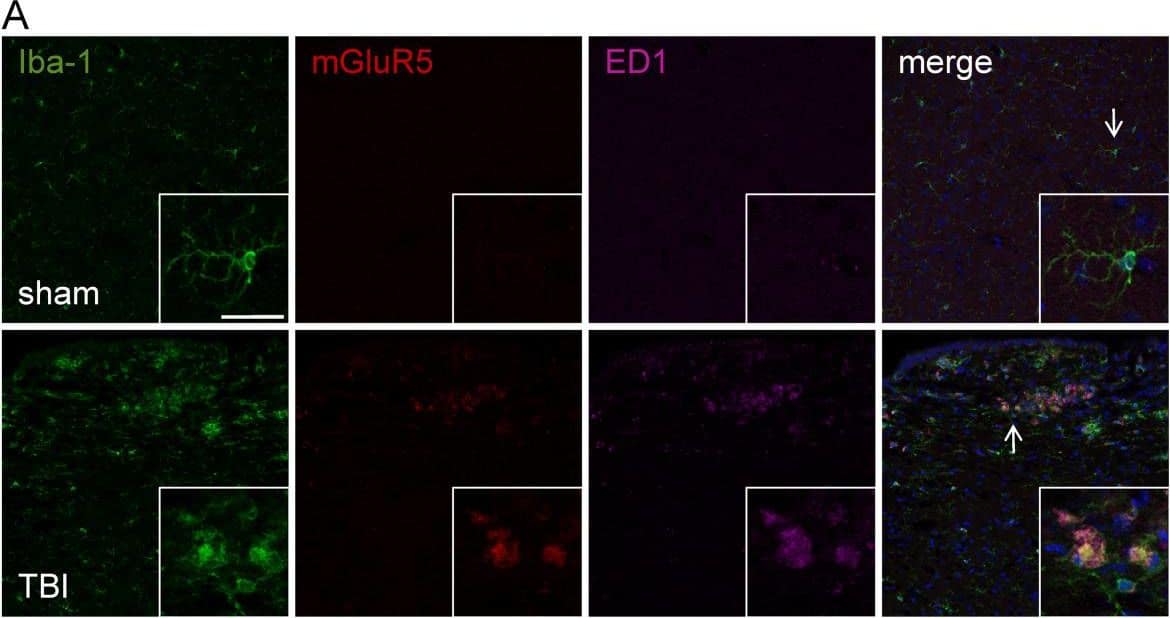
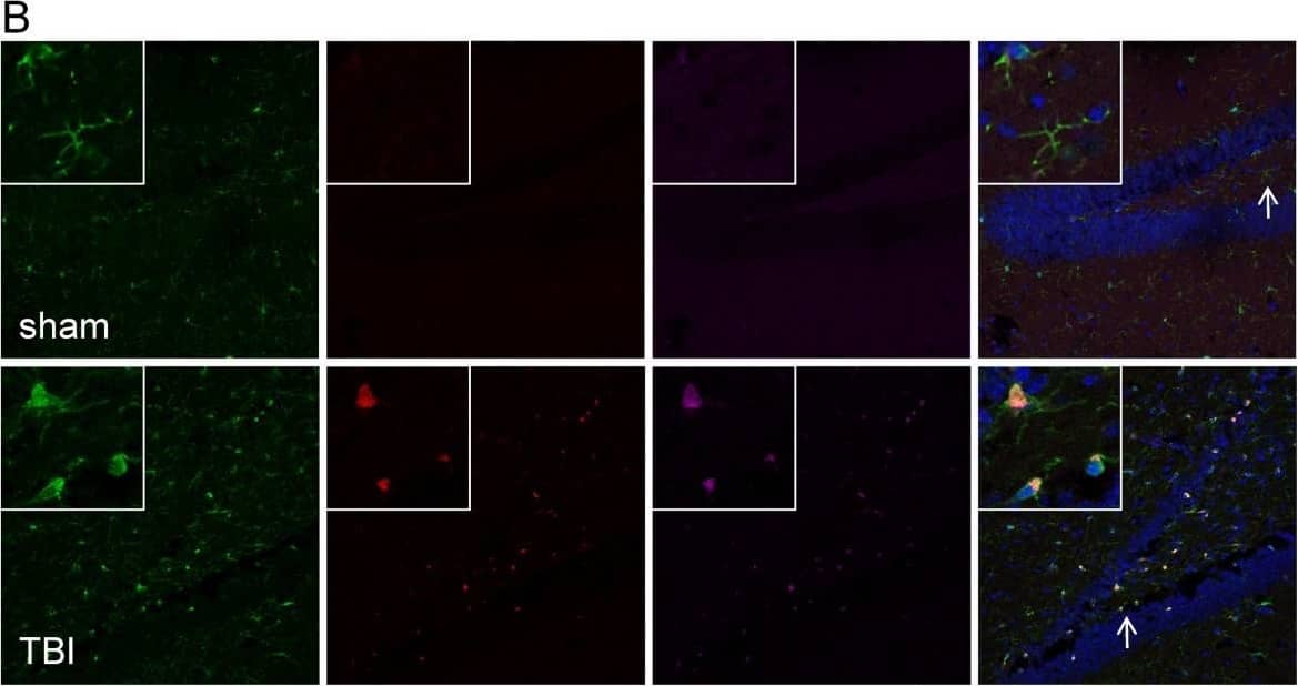
Detection of Mouse mGluR5 by Immunocytochemistry/Immunofluorescence mGluR5 is expressed in chronically activated microglia at one month post TBI. (A and B) mGluR5 expression was evaluated in the cortex (A) and hippocampus (B) of sham or TBI brains at one month post-injury. Iba-1 (green) and ED1 (magenta) labeled activated microglia. mGluR5 expression (red) was undetectable in resting microglia that displayed a ramified cellular morphology, but was strongly up-regulated in highly reactive microglia that displayed a hypertrophic or bushy cellular morphology and co-expressed ED1 (merged). (C) The membrane bound component of the NADPH oxidase enzyme, gp91phox (red), co-localized (merged) with ED1-positive reactive microglial (green) at one month post-TBI. Bar = 25 μm. Image collected and cropped by CiteAb from the following publication (https://pubmed.ncbi.nlm.nih.gov/22373400), licensed under a CC-BY license. Not internally tested by R&D Systems.
 View Larger
Detection of Human mGluR5 by Immunocytochemistry/Immunofluorescence mGluR5 is expressed in chronically activated microglia at one month post TBI. (A and B) mGluR5 expression was evaluated in the cortex (A) and hippocampus (B) of sham or TBI brains at one month post-injury. Iba-1 (green) and ED1 (magenta) labeled activated microglia. mGluR5 expression (red) was undetectable in resting microglia that displayed a ramified cellular morphology, but was strongly up-regulated in highly reactive microglia that displayed a hypertrophic or bushy cellular morphology and co-expressed ED1 (merged). (C) The membrane bound component of the NADPH oxidase enzyme, gp91phox (red), co-localized (merged) with ED1-positive reactive microglial (green) at one month post-TBI. Bar = 25 μm. Image collected and cropped by CiteAb from the following publication (https://pubmed.ncbi.nlm.nih.gov/22373400), licensed under a CC-BY license. Not internally tested by R&D Systems.
Reconstitution Calculator
Preparation and Storage
- 12 months from date of receipt, -20 to -70 °C as supplied.
- 1 month, 2 to 8 °C under sterile conditions after reconstitution.
- 6 months, -20 to -70 °C under sterile conditions after reconstitution.
Background: mGluR5
Human metabotropic glutamate receptor 5 (mGluR5; also known as mGluR5b) is a 150 kDa, 7-transmembrane glycoprotein that belongs to group I of the C-family of G-protein coupled receptors. mGluR5 is constitutively expressed and regulates neuronal ion channel activity. Human mGluR5 is 1212 amino acids (aa) in length and contains an N-terminal extracellular domain (ECD) of 558 aa. Through its ECD, mGluR5 either homodimerizes or heterodimerizes with the Ca++-sensor receptor. There is one alternate splice form (mGluR5a) that shows a 32 aa deletion between aa 877-908 in the cytoplasmic tail. Over aa 21-509, human mGluR5 shares 98% aa sequence identity with mouse, rat, and dog mGluR5.
Product Datasheets
Citations for Human mGluR5 Antibody
R&D Systems personnel manually curate a database that contains references using R&D Systems products. The data collected includes not only links to publications in PubMed, but also provides information about sample types, species, and experimental conditions.
2
Citations: Showing 1 - 2
Filter your results:
Filter by:
-
The role of metabotropic glutamate receptor 5 on the stromal cell-derived factor-1/CXCR4 system in oral cancer.
Authors: Kuribayashi, Nobuyuki, Uchida, Daisuke, Kinouchi, Makoto, Takamaru, Natsumi, Tamatani, Tetsuya, Nagai, Hirokazu, Miyamoto, Youji
PLoS ONE, 2013-11-13;8(11):e80773.
Species: Human
Sample Types: Whole Cells
Applications: Flow Cytometry -
Delayed mGluR5 activation limits neuroinflammation and neurodegeneration after traumatic brain injury.
Authors: Byrnes KR, Loane DJ, Stoica BA
J Neuroinflammation, 2012-02-28;9(0):43.
Species: Mouse
Sample Types: Whole Tissue
Applications: IHC-P
FAQs
No product specific FAQs exist for this product, however you may
View all Antibody FAQsReviews for Human mGluR5 Antibody
There are currently no reviews for this product. Be the first to review Human mGluR5 Antibody and earn rewards!
Have you used Human mGluR5 Antibody?
Submit a review and receive an Amazon gift card.
$25/€18/£15/$25CAN/¥75 Yuan/¥2500 Yen for a review with an image
$10/€7/£6/$10 CAD/¥70 Yuan/¥1110 Yen for a review without an image


