Human MMR/CD206 Antibody Summary
Leu19-Lys1383 (Thr399Ala) & (Leu407Phe),
Accession # P22897
Applications
Please Note: Optimal dilutions should be determined by each laboratory for each application. General Protocols are available in the Technical Information section on our website.
Scientific Data
 View Larger
Detection of Human MMR/CD206 by Western Blot. Western blot shows lysates of human immature dendritic cells. PVDF Membrane was probed with 1 µg/mL of Mouse Anti-Human MMR/CD206 Monoclonal Antibody (Catalog # MAB25341) followed by HRP-conjugated Anti-Mouse IgG Secondary Antibody (Catalog # HAF007). A specific band was detected for MMR/CD206 at approximately 150 kDa (as indicated). This experiment was conducted under reducing conditions and using Immunoblot Buffer Group 1.
 View Larger
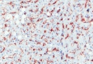
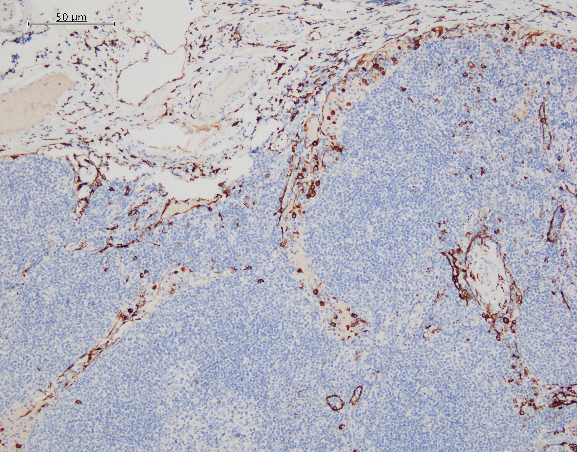
MMR/CD206 in Human Liver. MMR/CD206 was detected in immersion fixed paraffin-embedded sections of human liver using Mouse Anti-Human MMR/CD206 Monoclonal Antibody (Catalog # MAB25341) at 15 µg/mL overnight at 4 °C. Before incubation with the primary antibody, tissue was subjected to heat-induced epitope retrieval using Antigen Retrieval Reagent-Basic (Catalog # CTS013). Tissue was stained using the Anti-Mouse HRP-DAB Cell & Tissue Staining Kit (brown; Catalog # CTS002) and counterstained with hematoxylin (blue). Specific staining was localized to endothelial cells in sinusoids. View our protocol for Chromogenic IHC Staining of Paraffin-embedded Tissue Sections.
 View Larger
Detection of Human MMR/CD206 by Simple WesternTM. Simple Western lane view shows lysates of human immature dendritic cells, loaded at 0.2 mg/mL. A specific band was detected for MMR/CD206 at approximately 228 kDa (as indicated) using 20 µg/mL of Mouse Anti-Human MMR/CD206 Monoclonal Antibody (Catalog # MAB25341). This experiment was conducted under reducing conditions and using the 66-440 kDa separation system.
Reconstitution Calculator
Preparation and Storage
- 12 months from date of receipt, -20 to -70 °C as supplied.
- 1 month, 2 to 8 °C under sterile conditions after reconstitution.
- 6 months, -20 to -70 °C under sterile conditions after reconstitution.
Background: MMR/CD206
The human Macrophage Mannose Receptor (MMR), also known as CD206 and MRC1 (mannose receptor C, type 1), is a 190 kDa scavenger receptor that is expressed on tissue macrophages, myeloid dendritic cells, and liver and lymphatic endothelial cells (1). It belongs to a family of receptors sharing similar protein structure that also includes DEC205, phospholipase A2 receptor, and Endo180 (2, 3). The human MMR protein is synthesized as a 1456 amino acid (aa) precursor that contains an 18 aa signal sequence, a 1371 aa extracellular region, a 21 aa transmembrane segment and a 46 aa cytoplasmic domain (4). Its extracellular region is composed of an N-terminal cysteine-rich domain, followed by a single fibronectin type II repeat, and eight C-type lectin carbohydrate recognition domains (CRD) (3, 4). Human and mouse MMR extracellular regions share 82% aa identity. The cysteine-rich domain mediates recognition of sulfated N-acetylgalactosamine, which occurs on some extracellular matrix proteins and is the terminal sugar of the unusual oligosaccharides present on pituitary hormones such as lutropin and thyrotropin (5). Several of the CRDs participate in the Ca2+-dependent recognition of carbohydrates showing a preference for branched sugars with terminal mannose, fucose or N‑acetylglucosamine (6). The cytoplasmic domain of MMR includes a tyrosine-based motif for internalization in clathrin-coated vesicles. Once internalized, ligands are released following acidification of phagosomes or endosomes, and the receptor is recycled to the cell surface (3, 7). MMR mediates phagocytosis upon binding to target structures that occur on a variety of pathogenic microorganisms including Gram-negative and Gram-positive bacteria, yeasts, parasites, and mycobacteria. MMR also functions to maintain homeostasis through the endocytosis of potentially harmful glycoproteins associated with inflammation (2, 3).
- East, L. and C. Isake (2002) Biochim. Biophys. Acta 1572:364.
- Chieppa, M. et al. (2003) J. Immunol. 171:4552.
- Figdor, C. et al. (2002) Nat. Rev. Immunol. 2:77.
- Taylor, M. et al. (1990) J. Biol. Chem. 265:12156.
- Leteux, C. et al. (2000) J. Exp. Med. 191:1117.
- Martinez-Pomares, L. et al. (2001) Immunobiology 204:527.
- Feinberg, H. et al. (2000) J. Biol. Chem. 275:21539.
Product Datasheets
Citations for Human MMR/CD206 Antibody
R&D Systems personnel manually curate a database that contains references using R&D Systems products. The data collected includes not only links to publications in PubMed, but also provides information about sample types, species, and experimental conditions.
10
Citations: Showing 1 - 10
Filter your results:
Filter by:
-
Macrophage-Myofibroblast Transition Contributes to Myofibroblast Formation in Proliferative Vitreoretinal Disorders
Authors: Abu El-Asrar AM, De Hertogh G, Allegaert E et al.
International journal of molecular sciences
-
Specification of fetal liver endothelial progenitors to functional zonated adult sinusoids requires c-Maf induction
Authors: Jesus Maria Gómez-Salinero, Franco Izzo, Yang Lin, Sean Houghton, Tomer Itkin, Fuqiang Geng et al.
Cell Stem Cell
-
Macrophage Mannose Receptor CD206 Predicts Prognosis in Community-acquired Pneumonia
Authors: T Kazuo, Y Suzuki, K Yoshimura, H Yasui, M Karayama, H Hozumi, K Furuhashi, N Enomoto, T Fujisawa, Y Nakamura, N Inui, K Yokomura, T Suda
Sci Rep, 2019-12-10;9(1):18750.
Species: Human
Sample Types: Whole Tissue
Applications: IHC-P -
Regional Differences Between Perisynovial and Infrapatellar Adipose Tissue Depots and Their Response to Class II and III Obesity in Patients with OA
Authors: NS Harasymowi, ND Clement, A Azfer, R Burnett, DM Salter, AH Simpson
Arthritis & rheumatology (Hoboken, N.J.), 2017-06-10;0(0):.
Species: Human
Sample Types: Whole Tissue
Applications: IHC -
Macrophage Activation in Pediatric Nonalcoholic Fatty Liver Disease (NAFLD) Correlates with Hepatic Progenitor Cell Response via Wnt3a Pathway
Authors: Guido Carpino
PLoS ONE, 2016-06-16;11(6):e0157246.
Species: Human
Sample Types: Whole Tissue
Applications: IHC-P -
Upregulation of C/EBP alpha Inhibits Suppressive Activity of Myeloid Cells and Potentiates Antitumor Response in Mice and Patients with Cancer
Authors: Ayumi Hashimoto, Debashis Sarker, Vikash Reebye, Sheba Jarvis, Mikael H. Sodergren, Andrew Kossenkov et al.
Clinical Cancer Research
-
Myeloid HO-1 modulates macrophage polarization and protects against ischemia-reperfusion injury
Authors: Min Zhang, Kojiro Nakamura, Shoichi Kageyama, Akeem O. Lawal, Ke Wei Gong, May Bhetraratana et al.
JCI Insight
-
Senescent Tumor Cells Build a Cytokine Shield in Colorectal Cancer
Authors: Choi YW, Kim YH, Oh SY et al.
Advanced Science
-
IL-4/IL-13-mediated polarization of renal macrophages/dendritic cells to an M2a phenotype is essential for recovery from acute kidney injury.
Authors: Zhang Ming-Zhi, Wang Xin, Wang Yinqiu et al.
Kidney International
-
Macrophage Cyclooxygenase-2 Protects Against Development of Diabetic Nephropathy.
Authors: Wang Xin, Yao Bing, Wang Yinqiu et al.
Diabetes
FAQs
No product specific FAQs exist for this product, however you may
View all Antibody FAQsReviews for Human MMR/CD206 Antibody
Average Rating: 4.3 (Based on 3 Reviews)
Have you used Human MMR/CD206 Antibody?
Submit a review and receive an Amazon gift card.
$25/€18/£15/$25CAN/¥75 Yuan/¥2500 Yen for a review with an image
$10/€7/£6/$10 CAD/¥70 Yuan/¥1110 Yen for a review without an image
Filter by:
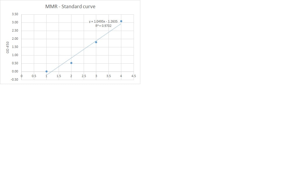
I used this antibody for developing a sandwich ELISA in combination with pAb (cat.AF2534) and protein (cat.2534 MR). This combination gives good standard curve but unfortunately did not detect any MMR in our samples.


