Human/Mouse BCAP Antibody Summary
Met462-Leu650
Accession # Q6ZUJ8
*Small pack size (-SP) is supplied either lyophilized or as a 0.2 µm filtered solution in PBS.
Applications
Please Note: Optimal dilutions should be determined by each laboratory for each application. General Protocols are available in the Technical Information section on our website.
Scientific Data
 View Larger
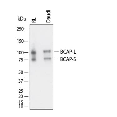
Detection of Human BCAP by Western Blot. Western Blot shows lysates of RL human non-Hodgkin's lymphoma B cell line and Daudi human Burkitt's lymphoma cell line. PVDF membrane was probed with 1 µg/ml of Goat Anti-Human/Mouse BCAP Antigen Affinity-purified Polyclonal Antibody (Catalog # AF4857) followed by HRP-conjugated Anti-Goat IgG Secondary Antibody (Catalog # HAF017). Specific bands were detected for BCAP at approximately 80 kDa and 100 kDa (as indicated). This experiment was conducted under reducing conditions and using Western Blot Buffer Group 1.
 View Larger
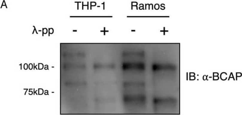
Detection of Human BCAP/PIK3AP1 by Western Blot BCAP is hyperphosphorylated in B-cell, macrophages, and Expi293F cells.A, lysates from THP-1 and Ramos cells were dephosphorylated with lambda -phosphatase and immunoblotted for BCAP. B, His-Avi-tagged BCAP expressed in Expi293F cells was purified and dephosphorylated with lambda -phosphatase before immunostaining for tyrosine and serine phosphorylation. C, phosphorylation sites of BCAP expressed in Expi293F cells were determined by phosphopeptide mapping. BCAP was digested with trypsin, chymotrypsin, Asp-N, and Glu-C prior to MS. Image collected and cropped by CiteAb from the following open publication (https://pubmed.ncbi.nlm.nih.gov/31527084), licensed under a CC-BY license. Not internally tested by R&D Systems.
Reconstitution Calculator
Preparation and Storage
- 12 months from date of receipt, -20 to -70 °C as supplied.
- 1 month, 2 to 8 °C under sterile conditions after reconstitution.
- 6 months, -20 to -70 °C under sterile conditions after reconstitution.
Background: BCAP
BCAP (B-cell adaptor for phosphoinositide 3-kinase (PI3K)) participates in linking the B cell antigen receptor (BCR) with the PI3K pathway. Tyrosine phosphorylation of BCAP by BCR-associated protein tyrosine kinases, such as Syk and Btk, generates binding sites for the p85 subunit of PI3K, resulting in activation of the PI3K pathway. Human and mouse BCAP contain 3 YXXM SH2 binding motifs for interaction with PI3K.
Product Datasheets
Citations for Human/Mouse BCAP Antibody
R&D Systems personnel manually curate a database that contains references using R&D Systems products. The data collected includes not only links to publications in PubMed, but also provides information about sample types, species, and experimental conditions.
2
Citations: Showing 1 - 2
Filter your results:
Filter by:
-
Negative Regulation of TLR Signaling by BCAP Requires Dimerization of Its DBB Domain
Authors: JU Lauenstein, MJ Scherm, A Udgata, MC Moncrieffe, DI Fisher, NJ Gay
J. Immunol., 2020-03-20;0(0):.
Species: Escherichia coli
Sample Types: Whole Cells
Applications: ICC -
Phosphorylation of the multi functional signal transducer B-cell adaptor protein (BCAP) promotes recruitment of multiple SH2/SH3 proteins including GRB2
Authors: JU Lauenstein, A Udgata, A Bartram, D De Sutter, DI Fisher, S Halabi, S Eyckerman, NJ Gay
J. Biol. Chem., 2019-09-16;0(0):.
Species: Human
Sample Types: Protein
Applications: Bioassay
FAQs
No product specific FAQs exist for this product, however you may
View all Antibody FAQsReviews for Human/Mouse BCAP Antibody
There are currently no reviews for this product. Be the first to review Human/Mouse BCAP Antibody and earn rewards!
Have you used Human/Mouse BCAP Antibody?
Submit a review and receive an Amazon gift card.
$25/€18/£15/$25CAN/¥75 Yuan/¥2500 Yen for a review with an image
$10/€7/£6/$10 CAD/¥70 Yuan/¥1110 Yen for a review without an image

