Human/Mouse Cathepsin L Antibody Summary
Glu113-Val333
Accession # P07711
Applications
Please Note: Optimal dilutions should be determined by each laboratory for each application. General Protocols are available in the Technical Information section on our website.
Scientific Data
 View Larger
Detection of Recombinant Human and Mouse Cathepsin L by Western Blot. Western blot shows 100 ng of Recombinant Human Cathepsin L (Catalog # 952-CY), Recombinant Mouse Cathepsin L (Catalog # 1515-CY), Recombinant Human Cathepsin V (Catalog # 1080-CY), Recombinant Human Cathepsin K, Recombinant Human Cathepsin S (Catalog # 1183-CY), Recombinant Human Cathepsin H (Catalog # 7516-CY), and Recombinant Human Cathepsin F. PVDF Membrane was probed with 1 µg/mL of Rat Anti-Human/Mouse Cathepsin L Monoclonal Antibody (Catalog # MAB9521) followed by HRP-conjugated Anti-Rat IgG Secondary Antibody (Catalog # HAF005). A specific band was detected for Cathepsin L at approximately 35 kDa (as indicated). This experiment was conducted under reducing conditions and using Immunoblot Buffer Group 3. For natural samples, we recommend the use of Catalog # AF1515.
 View Larger
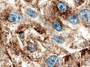
Cathepsin L in Human Kidney. Cathepsin L was detected in immersion fixed paraffin-embedded sections of human kidney using Rat Anti-Human/Mouse Cathepsin L Monoclonal Antibody (Catalog # MAB9521) at 5 µg/mL overnight at 4 °C. Tissue was stained using the Anti-Rat HRP-DAB Cell & Tissue Staining Kit (brown; Catalog # CTS017) and counterstained with hematoxylin (blue). Specific staining was localized to cytoplasm in tubular epithelial cells. View our protocol for Chromogenic IHC Staining of Paraffin-embedded Tissue Sections.
 View Larger
Cathepsin L in Mouse Kidney. Cathepsin L was detected in immersion fixed frozen sections of mouse kidney using Rat Anti-Human/Mouse Cathepsin L Monoclonal Antibody (Catalog # MAB9521) at 8 µg/mL overnight at 4 °C. Tissue was stained using the Anti-Rat HRP-DAB Cell & Tissue Staining Kit (brown; Catalog # CTS017) and counterstained with hematoxylin (blue). Specific staining was localized to cytoplasm in tubular epithelial cells. View our protocol for Chromogenic IHC Staining of Frozen Tissue Sections.
Preparation and Storage
- 12 months from date of receipt, -20 to -70 °C as supplied.
- 1 month, 2 to 8 °C under sterile conditions after reconstitution.
- 6 months, -20 to -70 °C under sterile conditions after reconstitution.
Background: Cathepsin L
Cathepsin L is a lysosomal cysteine protease expressed in most eukaryotic cells. Cathepsin L is known to hydrolyze a number of proteins, including the proform of urokinase-type plasminogen activator, which is activated by Cathepsin L cleavage (1). Cathepsin L has also been shown to proteolytically inactivate alpha 1-antitrypsin and secretory leucoprotease inhibitor, two major protease inhibitors of the respiratory tract (2). These observations, combined with the demonstration of increased Cathepsin L activity in the epithelial lining fluid of the lungs of emphysema patients, have led to the suggestion that the enzyme may be involved in the progression of this disease. Cathepsin L has also been identified as a major excreted protein of transformed fibroblasts, indicating the enzyme could be involved in malignant tumor growth (3).
- Goretzki, L. et al. (1992) FEBS Lett. 297:112.
- Taggart, C.C. et al. (2001) J. Biol. Chem. 276:33345.
- Gottesman, M.M. and F. Cabral (1981) Biochemistry 20:1659.
Product Datasheets
Citations for Human/Mouse Cathepsin L Antibody
R&D Systems personnel manually curate a database that contains references using R&D Systems products. The data collected includes not only links to publications in PubMed, but also provides information about sample types, species, and experimental conditions.
12
Citations: Showing 1 - 10
Filter your results:
Filter by:
-
cPLA2 activation contributes to lysosomal defects leading to impairment of autophagy after spinal cord injury
Authors: Y Li, JW Jones, H M C Choi, C Sarkar, MA Kane, EY Koh, MM Lipinski, J Wu
Cell Death Dis, 2019-07-11;10(7):531.
-
PLA2G4A/cPLA2-mediated lysosomal membrane damage leads to inhibition of autophagy and neurodegeneration after brain trauma
Authors: C Sarkar, JW Jones, N Hegdekar, JA Thayer, A Kumar, AI Faden, MA Kane, MM Lipinski
Autophagy, 2019-06-25;0(0):1-20.
Species: Mouse
Sample Types: Whole Tissue
Applications: IHC -
CCT complex restricts neuropathogenic protein aggregation via autophagy
Nat Commun, 2016-12-08;7(0):13821.
Species: Human
Sample Types: Cell Lysates
Applications: Western Blot -
A C9ORF72/SMCR8-containing complex regulates ULK1 and plays a dual role in autophagy
Sci Adv, 2016-09-02;2(9):e1601167.
Species: Mouse
Sample Types: Cell Lysates
Applications: Western Blot -
The PI3K regulatory subunits p55alpha and p50alpha regulate cell death in vivo.
Authors: Pensa S, Neoh K, Resemann H, Kreuzaler P, Abell K, Clarke N, Reinheckel T, Kahn C, Watson C
Cell Death Differ, 2014-06-06;21(9):1442-50.
Species: Mouse
Sample Types: Cell Lysates, Whole Tissue
Applications: IHC-P, Western Blot -
A 3-D in vitro co-culture model of mammary gland involution
Authors: Jonathan J. Campbell, Laur-Alexandru Botos, Timothy J. Sargeant, Natalia Davidenko, Ruth E. Cameron, Christine J. Watson
Integr Biol (Camb)
-
Macrophages are crucial for epithelial cell death and adipocyte repopulation during mammary gland involution.
Authors: O'Brien J, Martinson H, Durand-Rougely C
Development, 2011-11-30;139(2):269-75.
Species: Mouse
Sample Types: Tissue Homogenates
Applications: Western Blot -
Stat3 controls lysosomal-mediated cell death in vivo.
Authors: Kreuzaler PA, Staniszewska AD, Li W, Omidvar N, Kedjouar B, Turkson J, Poli V, Flavell RA, Clarkson RW, Watson CJ
Nat. Cell Biol., 2011-02-20;13(3):303-9.
Species: Mouse
Sample Types: Cell Lysates
Applications: Western Blot -
P2X(7) receptor-mediated release of cathepsins from macrophages is a cytokine-independent mechanism potentially involved in joint diseases.
Authors: Lopez-Castejon G, Theaker J, Pelegrin P
J. Immunol., 2010-07-16;185(4):2611-9.
Species: Human, Mouse
Sample Types: Cell Culture Supernates
Applications: Western Blot -
The cystatin M/E-cathepsin L balance is essential for tissue homeostasis in epidermis, hair follicles, and cornea.
Authors: Zeeuwen PL, van Vlijmen-Willems IM, Cheng T
FASEB J., 2010-05-21;24(10):3744-55.
Species: Mouse
Sample Types: Whole Tissue
Applications: IHC-P -
Primary tumour expression of the cysteine cathepsin inhibitor Stefin A inhibits distant metastasis in breast cancer.
Authors: Parker BS, Ciocca DR, Bidwell BN, Gago FE, Fanelli MA, George J, Slavin JL, Moller A, Steel R, Pouliot N, Eckhardt BL, Henderson MA, Anderson RL
J. Pathol., 2008-02-01;214(3):337-46.
Species: Mouse
Sample Types: Whole Tissue
Applications: IHC-P -
Cystatin M/E is a high affinity inhibitor of cathepsin V and cathepsin L by a reactive site that is distinct from the legumain-binding site. A novel clue for the role of cystatin M/E in epidermal cornification.
Authors: Cheng T, Hitomi K, van Vlijmen-Willems IM, de Jongh GJ, Yamamoto K, Nishi K, Watts C, Reinheckel T, Schalkwijk J, Zeeuwen PL
J. Biol. Chem., 2006-03-24;281(23):15893-9.
Species: Human
Sample Types: Whole Tissue
Applications: IHC
FAQs
No product specific FAQs exist for this product, however you may
View all Antibody FAQsReviews for Human/Mouse Cathepsin L Antibody
Average Rating: 4.8 (Based on 4 Reviews)
Have you used Human/Mouse Cathepsin L Antibody?
Submit a review and receive an Amazon gift card.
$25/€18/£15/$25CAN/¥75 Yuan/¥2500 Yen for a review with an image
$10/€7/£6/$10 CAD/¥70 Yuan/¥1110 Yen for a review without an image
Filter by:


