Human/Mouse CELSR2 Antibody
Human/Mouse CELSR2 Antibody Summary
Cys51-Phe231
Accession # Q9HCU4
Applications
Please Note: Optimal dilutions should be determined by each laboratory for each application. General Protocols are available in the Technical Information section on our website.
Scientific Data
 View Larger
Detection of CELSR2 by Western Blot. Western blot shows lysates of HEK293 human embryonic kidney cell line either mock transfected or transfected with human CELSR2. PVDF Membrane was probed with 1 µg/mL of Goat Anti-Human CELSR2 Antigen Affinity-purified Polyclonal Antibody (Catalog # AF6739) followed by HRP-conjugated Anti-Goat IgG Secondary Antibody (Catalog # HAF019). A specific band was detected for CELSR2 at approximately 240 kDa (as indicated). This experiment was conducted under reducing conditions and using Immunoblot Buffer Group 8.
 View Larger
Detection of CELSR2 in SH‑SY5Y Human Cell Line by Flow Cytometry. SH-SY5Y human neuroblastoma cell line was stained with Goat Anti-Human CELSR2 Antigen Affinity-purified Polyclonal Antibody (Catalog # AF6739, filled histogram) or isotype control antibody (Catalog # AB-108-C, open histogram), followed by Allophycocyanin-conjugated Anti-Goat IgG Secondary Antibody (Catalog # F0108).
 View Larger
Detection of CELSR2 in bEnd.3 Mouse Cell Line by Flow Cytometry. bEnd.3 mouse endothelioma cell line was stained with Goat Anti-Human CELSR2 Antigen Affinity-purified Polyclonal Antibody (Catalog # AF6739, filled histogram) or isotype control antibody (Catalog # AB-108-C, open histogram), followed by Allophycocyanin-conjugated Anti-Goat IgG Secondary Antibody (Catalog # F0108).
 View Larger
CELSR2 in Human Breast. CELSR2 was detected in immersion fixed paraffin-embedded sections of human breast using Goat Anti-Human CELSR2 Antigen Affinity-purified Polyclonal Antibody (Catalog # AF6739) at 10 µg/mL overnight at 4 °C. Tissue was stained using the Anti-Goat HRP-DAB Cell & Tissue Staining Kit (brown; Catalog # CTS008) and counterstained with hematoxylin (blue). Lower panel shows a lack of labeling when primary antibodies are omitted and tissue is stained only with secondary antibody followed by incubation with detection reagents. Specific staining was localized to ductal epithelium. View our protocol for Chromogenic IHC Staining of Paraffin-embedded Tissue Sections.
Reconstitution Calculator
Preparation and Storage
- 12 months from date of receipt, -20 to -70 °C as supplied.
- 1 month, 2 to 8 °C under sterile conditions after reconstitution.
- 6 months, -20 to -70 °C under sterile conditions after reconstitution.
Background: CELSR2
CELSR2 (Cadherin EGF LAG seven-pass G-type receptor 2; also cadherin family member 10/CDHF10, Flamingo1 and EGFL2) is a 300-330 kDa member of the LN‑7TM subfamily, GPCR 2 family of proteins. It is expressed on neurons, breast epithelium, Sertoli cells and germ cells, and through homophilic interactions, serves as either an adhesion or guidance molecule. Mature human CELSR2 is 2892 amino acids in length (aa 32-2923). It is a highly complex 7-transmembrane protein that contains a 2349 aa extended N-terminal extracellular region (aa 32-2380) plus a 310 aa C-terminal cytoplasmic domain. The N-terminal region contains nine consecutive cadherin domains (aa 182-1146) followed by a mixture of seven EGF-like and three laminin-like domains. There is a proteolytic cleavage site between Met2356-Thr2357 that generates a 250 kDa soluble fragment and a (mature) 60-65 kDa transmembrane segment that may reside on the cell membrane. Over aa 51‑231, human CELSR2 shares 93% aa identity with mouse CELSR2.
Product Datasheets
Citations for Human/Mouse CELSR2 Antibody
R&D Systems personnel manually curate a database that contains references using R&D Systems products. The data collected includes not only links to publications in PubMed, but also provides information about sample types, species, and experimental conditions.
2
Citations: Showing 1 - 2
Filter your results:
Filter by:
-
SNX27 Deletion Causes Hydrocephalus by Impairing Ependymal Cell Differentiation and Ciliogenesis
Authors: Xin Wang
J. Neurosci, 2016-12-14;36(50):12586-12597.
Species: Mouse
Sample Types: Tissue Homogenates
Applications: Western Blot -
Celsr1 and Celsr2 exhibit distinct adhesive interactions and contributions to planar cell polarity
Authors: Lena P. Basta, Parijat Sil, Rebecca A. Jones, Katherine A. Little, Gabriela Hayward-Lara, Danelle Devenport
Frontiers in Cell and Developmental Biology
FAQs
No product specific FAQs exist for this product, however you may
View all Antibody FAQsReviews for Human/Mouse CELSR2 Antibody
Average Rating: 4 (Based on 1 Review)
Have you used Human/Mouse CELSR2 Antibody?
Submit a review and receive an Amazon gift card.
$25/€18/£15/$25CAN/¥75 Yuan/¥2500 Yen for a review with an image
$10/€7/£6/$10 CAD/¥70 Yuan/¥1110 Yen for a review without an image
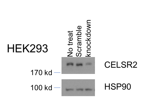
Filter by:

