Human/Mouse MSX1 Antibody Summary
Met1-Thr165
Accession # P28360
Applications
Please Note: Optimal dilutions should be determined by each laboratory for each application. General Protocols are available in the Technical Information section on our website.
Scientific Data
 View Larger
Detection of Human MSX1 by Western Blot. Western blot shows lysates of HeLa human cervical epithelial carcinoma cell line. Gels were loaded with 30 µg of whole cell lysate (WCL), 20 µg of cytoplasmic (Cyto), and 10 µg of nuclear extracts (Nuc). PVDF membrane was probed with 0.1 µg/mL Goat Anti-Human/Mouse MSX1 Antigen Affinity-purified Polyclonal Antibody (Catalog # AF5045) followed by HRP-conjugated Anti-Goat IgG Secondary Antibody (Catalog # HAF017). A specific band for MSX1 was detected at approximately 40 kDa (as indicated). This experiment was conducted under reducing conditions and using Immunoblot Buffer Group 1.
 View Larger
MSX1 in Human Ovarian Cancer Tissue. MSX1 was detected in immersion fixed paraffin-embedded sections of human ovarian cancer tissue using Goat Anti-Human/Mouse MSX1 Antigen Affinity-purified Polyclonal Antibody (Catalog # AF5045) at 0.3, 1.0 and 3.0 µg/mL overnight at 4 °C. Tissue was stained using the Anti-Goat HRP-DAB Cell & Tissue Staining Kit (brown; Catalog # CTS008) and counterstained with hematoxylin (blue). Specific staining was localized to nuclei. View our protocol for Chromogenic IHC Staining of Paraffin-embedded Tissue Sections.
 View Larger
Detection of Human MSX1 by Western Blot Regulation of MSX1 expression is mediated by pSMAD1/5/8 in the human cap-stage tooth germs. (A–C) Immunostaining shows that MSX1 expression is abundant in dimethyl sulfoxide (DMSO)-treated human cap-stage tooth germs, dramatically reduced in dorsomorphin treated human cap-stage tooth germ, and unaltered in U0126 + SB203580 + SP600125 treated human cap-stage tooth germs. (D) A Western blot assay confirms the dramatically reduced expression of MSX1 in dorsomorphin treated human cap-stage tooth germs. Actin was used as the internal control. (E) Quantitative analysis of the Western blot assay. de, the dental epithelium; dm, the dental mesenchyme. Error bars represent SD. ***p < 0.001. Bar = 50 μm. Image collected and cropped by CiteAb from the following publication (https://pubmed.ncbi.nlm.nih.gov/35211032), licensed under a CC-BY license. Not internally tested by R&D Systems.
Reconstitution Calculator
Preparation and Storage
- 12 months from date of receipt, -20 to -70 °C as supplied.
- 1 month, 2 to 8 °C under sterile conditions after reconstitution.
- 6 months, -20 to -70 °C under sterile conditions after reconstitution.
Background: MSX1
MSX1 (Msh homeobox homology 1) is a member of the muscle segment homoebox gene family. MSX1 is involved in limb-pattern formation, craniofacial development, odontogenesis, and tumor growth inhibition. MSX1 functions as a transcriptional repressor. MSX1 has been shown to interact with the linker histone, H1B, and repress transcription of the MyoD promoter. Chromosomal abnormalities involving MSX1 have been associated with the Wolf-Hirschhorn syndrome characterized by heart defects and mental retardation.
Product Datasheets
Citations for Human/Mouse MSX1 Antibody
R&D Systems personnel manually curate a database that contains references using R&D Systems products. The data collected includes not only links to publications in PubMed, but also provides information about sample types, species, and experimental conditions.
11
Citations: Showing 1 - 10
Filter your results:
Filter by:
-
Endothelial Msx1 transduces hemodynamic changes into an arteriogenic remodeling response
Authors: Ine Vandersmissen, Sander Craps, Maarten Depypere, Giulia Coppiello, Nick van Gastel, Frederik Maes et al.
Journal of Cell Biology
-
Differential Expression of NF2 in Neuroepithelial Compartments Is Necessary for Mammalian Eye Development.
Authors: Moon K H, Kim H T et al.
Dev Cell
-
Conditional deletion of Bmp2 in cranial neural crest cells recapitulates Pierre Robin sequence in mice
Authors: Yixuan Chen, Zhengsen Wang, YiPing Chen, Yanding Zhang
Cell and Tissue Research
-
SOX9-positive pituitary stem cells differ according to their position in the gland and maintenance of their progeny depends on context
Authors: Rizzoti, K;Chakravarty, P;Sheridan, D;Lovell-Badge, R;
Science advances
Species: Mouse
Sample Types: Whole Tissue
Applications: IHC -
Generation of a MSX1 knockout human embryonic stem cell line using CRISPR/Cas9 technology
Authors: W Chiu, A Li, T Wang, W Li, X Zhang
Stem Cell Research, 2022-02-26;60(0):102729.
Species: Human
Sample Types: Whole Cells
Applications: ICC -
Operation of the Atypical Canonical Bone Morphogenetic Protein Signaling Pathway During Early Human Odontogenesis
Authors: X Hu, C Lin, N Ruan, Z Huang, Y Zhang, X Hu
Frontiers in Physiology, 2022-02-08;13(0):823275.
Species: Human
Sample Types: Cell Lysates, Whole Cells, Whole Tissue
Applications: ICC, IHC, Western Blot -
Lef1 restricts ectopic crypt formation and tumor cell growth in intestinal adenomas
Authors: S Heino, S Fang, M Lähde, J Högström, S Nassiri, A Campbell, D Flanagan, A Raven, M Hodder, N Nasreddin, HH Xue, M Delorenzi, S Leedham, TV Petrova, O Sansom, K Alitalo
Science Advances, 2021-11-17;7(47):eabj0512.
Species: Mouse
Sample Types: Whole Tissue
Applications: IHC -
SARS-CoV-2 Infects the Brain Choroid Plexus and Disrupts the Blood-CSF Barrier in Human Brain Organoids
Authors: L Pellegrini, A Albecka, DL Mallery, MJ Kellner, D Paul, AP Carter, LC James, MA Lancaster
Cell Stem Cell, 2020-10-13;0(0):.
Species: Human
Sample Types: Organoid
Applications: IHC -
Msx1 loss suppresses formation of the ectopic crypts developed in the Apc-deficient small intestinal epithelium
Authors: M Horazna, L Janeckova, J Svec, O Babosova, D Hrckulak, M Vojtechova, K Galuskova, E Sloncova, M Kolar, H Strnad, V Korinek
Sci Rep, 2019-02-07;9(1):1629.
Species: Human, Mouse
Sample Types: Cell Lysates, Whole Tissue
Applications: IHC-P, Western Blot -
A new strategy to measure intercellular adhesion forces in mature cell-cell contacts
Authors: A Sancho, I Vandersmis, S Craps, A Luttun, J Groll
Sci Rep, 2017-04-10;7(0):46152.
Species: Human
Sample Types: Cell Lysates, Whole Cells
Applications: ICC, Western Blot -
Unraveling a novel transcription factor code determining the human arterial-specific endothelial cell signature.
Authors: Aranguren X, Agirre X, Beerens M, Coppiello G, Uriz M, Vandersmissen I, Benkheil M, Panadero J, Aguado N, Pascual-Montano A, Segura V, Prosper F, Luttun A
Blood, 2013-10-09;122(24):3982-92.
FAQs
No product specific FAQs exist for this product, however you may
View all Antibody FAQsReviews for Human/Mouse MSX1 Antibody
Average Rating: 4.7 (Based on 3 Reviews)
Have you used Human/Mouse MSX1 Antibody?
Submit a review and receive an Amazon gift card.
$25/€18/£15/$25CAN/¥75 Yuan/¥2500 Yen for a review with an image
$10/€7/£6/$10 CAD/¥70 Yuan/¥1110 Yen for a review without an image
Filter by:
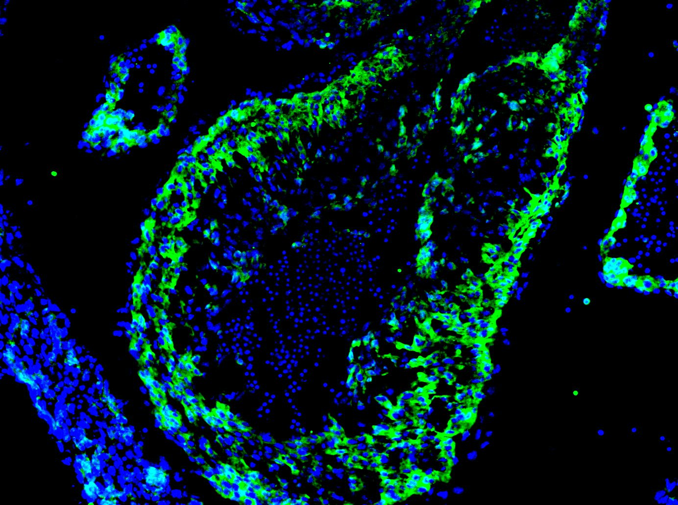
Fixed 4% PFA overnight.
Blocked with 1% BSA
Primary antibody dilution - 1:20
Secondary antibody - Invitrogen Alexa Fluor 488
Secondary antibody dilution - 1:1000
Stained on a E12.5 mouse heart section.
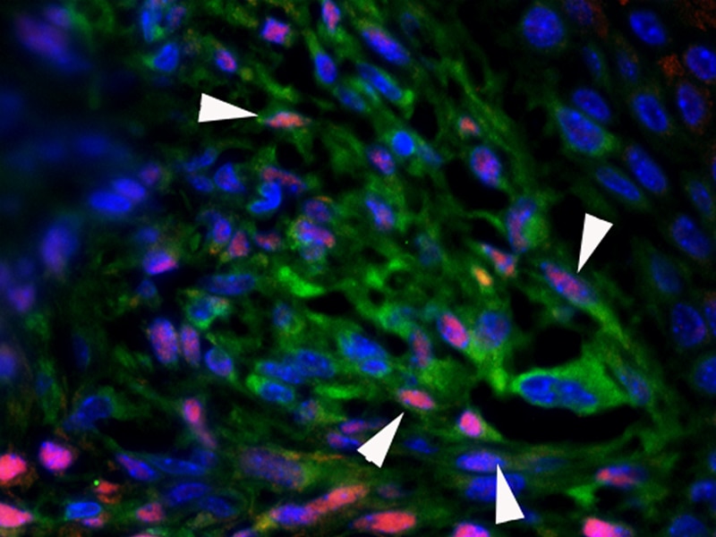
Primary antibody was used at 1:100 and incubated at 4°C overnight.
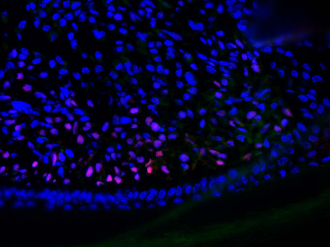
Msx1 expression on frozen sections of regenerating mouse digit was detected with AF5045 (1:200) incubated overnight in fridge ( 4°C) in PBS/3% BSA/0.1% Triton-X100, secondary antibody (1:500) incubated at room temperature for 90 minutes, followed by nuclei counterstaining with DAPI.


