Human/Mouse PARP Antibody Summary
Val71-Pro329
Accession # NP_031441
Applications
Please Note: Optimal dilutions should be determined by each laboratory for each application. General Protocols are available in the Technical Information section on our website.
Scientific Data
 View Larger
Detection of Human PARP by Western Blot. Western blot shows lysates of Jurkat human acute T cell leukemia cell line untreated (-) or treated with anti-Fas and 1 µM staurosporine (STS) for 0 to 4 hours (h; as indicated). PVDF membrane was probed with 0.6 µg/mL of Rat Anti-Human/Mouse PARP Monoclonal Antibody (Catalog # MAB600) followed by HRP-conjugated Anti-Rat IgG Secondary Antibody (Catalog # HAF005). Specific bands were detected for PARP at approximately 116 kDa and the 84 kDa product of PARP cleavage (as indicated). This experiment was conducted under reducing conditions and using Immunoblot Buffer Group 2.
 View Larger
Detection of Mouse PARP by Western Blot. Western blot shows lysates of NIH-3T3 mouse embryonic fibroblast cell line treated with 1 µM staurosporine (STS) for 0 to 4 hours (h; as indicated). PVDF membrane was probed with 0.6 µg/mL of Rat Anti-Human/Mouse PARP Monoclonal Antibody (Catalog # MAB600) followed by HRP-conjugated Anti-Rat IgG Secondary Antibody (Catalog # HAF005). Specific bands were detected for PARP at approximately 116 kDa and the 60-70 kDa bands generated with staurosporine treatment (as indicated). This experiment was conducted under reducing conditions and using Immunoblot Buffer Group 2.
 View Larger
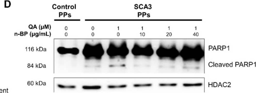
Detection of Human PARP by Western Blot The treatment of n-BP rescued the progression of QA-induced excitotoxicity in SCA3 PPs. The control and/or SCA3 PPs were treated with or without QA (1 μM) in the presence of n-BP (0, 10, 20, and 40 μg/mL) for 12 h. (A) Images of QA-treated SCA3 PPs in the presence of BP (40 μg/mL). (B) Confocal images of poly Q localization in BP (40 μg/mL)-treated SCA3 PPs in the presence of QA (1 um). When compared with Figure 2C, 40 μg/mL of n-BP prevented the amounts of colocalized polyQ in the cell nucleus of PPs. The distribution of polyQ in the nucleus is indicated by white arrowheads. Scale bar = 100 μm; (C) protein analysis of wild-type, mutant ATXN3 and its proteolytic fragment in cell lysates of control and SCA3 PPs, as assessed by immunoblots. Soluble, soluble protein; Insoluble, insoluble protein; (D,E) representative immunoblot of control and cleaved PARP1 in cell lysates of control and SCA3 PPs, as assessed by immunoblots. The HDAC2 were used as loading controls. The quantification from three independent images is presented as the means ± standard deviation. t(4) = 2.824, p < 0.05, for lane 1 vs. lane 2; t(4) = 5.613, p < 0.01, for lane 1 vs. lane 3; t(4) = 3.522, p < 0.05, for lane 1 vs. lane 4; (F) the quantification result of calcium concentration in SCA3 PPs, as assessed by Fura-2 indicator. Cells were treated with QA (1 μM) in the presence of n-BP (0, 10, 20, and 40 μg/mL). Data are presented as the means ± standard deviation. t(4) = 2.95, p < 0.05; (G) the calpain activity in control and SCA3 PPs cell lysates, as assessed by ELISA assay. The quantification results from three independent replicates are presented as the means ± standard deviation. t(4) = 3.27, p < 0.05, for lane 3 vs. lane 4; t(4) = 17.55, p < 0.01, for lane 3 vs. lane 5; t(4) = 9.019, p < 0.01, for lane 3 vs. lane 6. (H) Protein analysis of calpain 1, calpain 2 and calpastatin in cell lysates of SCA3 PPs, as assessed by immunoblots. Cells were treated with or without QA (1 μM) in the presence of n-BP (0, 10, 20, and 40 μg/mL). *, p < 0.05; **, p < 0.01. Image collected and cropped by CiteAb from the following publication (https://pubmed.ncbi.nlm.nih.gov/35163312), licensed under a CC-BY license. Not internally tested by R&D Systems.
Reconstitution Calculator
Preparation and Storage
- 12 months from date of receipt, -20 to -70 °C as supplied.
- 1 month, 2 to 8 °C under sterile conditions after reconstitution.
- 6 months, -20 to -70 °C under sterile conditions after reconstitution.
Background: PARP
PARP [Poly(ADP-ribose) Polymerase], also known as ADPRT and PPOL, is a 118-kDa enzyme that uses NAD as a substrate to catalyze the covalent transfer of ADP-ribose to a variety of nuclear protein acceptors. ADP ribosyltransferase is required for cellular repair, and PARP expression is induced by single-strand breaks in DNA. PARP is proteolytically cleaved by Caspase-3 into two fragments of 89- and 24-kDa in one of the hallmark events of apoptosis.
Product Datasheets
Citations for Human/Mouse PARP Antibody
R&D Systems personnel manually curate a database that contains references using R&D Systems products. The data collected includes not only links to publications in PubMed, but also provides information about sample types, species, and experimental conditions.
6
Citations: Showing 1 - 6
Filter your results:
Filter by:
-
TET-mediated DNA hydroxymethylation is negatively influenced by the PARP-dependent PARylation
Authors: A Toli?, M Ravichandr, J Raji?, M ?or?evi?, M ?or?evi?, S Dini?, N Grdovi?, JA Jovanovi?, M Mihailovi?, N Nestorovi?, TP Jurkowski, AS Uskokovi?, MS Vidakovi?
Epigenetics & Chromatin, 2022-04-05;15(1):11.
Species: Mouse
Sample Types: Whole Cells
Applications: IHC -
The Molecular Basis of Tight Nuclear Tethering and Inactivation of cGAS
Authors: B Zhao, P Xu, CM Rowlett, T Jing, O Shinde, Y Lei, AP West, WR Liu, P Li
Nature, 2020-09-10;0(0):.
Species: Human
Sample Types: Cell Lysates, Whole Cells
Applications: ICC, Western Blot -
RNA stability regulates human T cell leukemia virus type 1 gene expression in chronically-infected CD4 T cells
Authors: HC Lin, PJ Simon, RM Ysla, SL Zeichner, G Brewer, AB Rabson
Virology, 2017-05-04;508(0):7-17.
Species: Human
Sample Types: Cell Lysates
Applications: Western Blot -
Conditional deletion of Nbs1 in murine cells reveals its role in branching repair pathways of DNA double-strand breaks.
Authors: Yang YG, Saidi A, Frappart PO, Barrucand C, Dumon-Jones V, Michelon J, Herceg Z
EMBO J., 2006-11-02;25(23):5527-38.
Species: Mouse
Sample Types: Cell Lysates
Applications: Western Blot -
Distinct promoters mediate constitutive and inducible Bcl-XL expression in malignant lymphocytes.
Authors: Habens F, Lapham AS, Dallman CL, Pickering BM, Michels J, Marcusson EG, Johnson PW, Packham G
Oncogene, 2006-09-18;26(13):1910-9.
Species: Human
Sample Types: Cell Lysates
Applications: Western Blot -
Transcriptional repressor activating transcription factor 3 protects human umbilical vein endothelial cells from tumor necrosis factor-alpha-induced apoptosis through down-regulation of p53 transcription.
Authors: Kawauchi J, Zhang C, Nobori K, Hashimoto Y, Adachi MT, Noda A, Sunamori M, Kitajima S
J. Biol. Chem., 2002-08-02;277(41):39025-34.
Species: Human
Sample Types: Whole Cells
Applications: Western Blot
FAQs
No product specific FAQs exist for this product, however you may
View all Antibody FAQsReviews for Human/Mouse PARP Antibody
Average Rating: 5 (Based on 1 Review)
Have you used Human/Mouse PARP Antibody?
Submit a review and receive an Amazon gift card.
$25/€18/£15/$25CAN/¥75 Yuan/¥2500 Yen for a review with an image
$10/€7/£6/$10 CAD/¥70 Yuan/¥1110 Yen for a review without an image
Filter by:

