Human/Mouse/Rat Akt2 Antibody Summary
Accession # P31751
Applications
Please Note: Optimal dilutions should be determined by each laboratory for each application. General Protocols are available in the Technical Information section on our website.
Scientific Data
 View Larger
Detection of Human/Mouse/Rat Akt2 by Western Blot. Western blot shows lysates of MCF-7 human breast cancer cell line, MDA-MB-468 human breast cancer cell line, and U937 human histiocytic lymphoma cell line. PVDF membrane was probed with 0.5 µg/mL Goat Anti-Human/Mouse/Rat Akt2 Antigen Affinity-purified Polyclonal Antibody (Catalog # AF23151) followed by HRP-conjugated Anti-Goat IgG Secondary Antibody (Catalog # HAF109). For additional reference, Recombinant Human Active Akt1 (Catalog # 1775-KS), recombinant human Akt2, and recombinant human Akt3 (5 ng/lane) were included. A specific band for Akt2 was detected at approximately 60 kDa (as indicated). This experiment was conducted under reducing conditions and using Immunoblot Buffer Group 1.
 View Larger
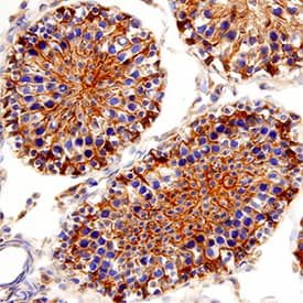
Akt2 in Rat Testis. Akt2 was detected in perfusion fixed frozen sections of rat testis using Goat Anti-Human/Mouse/Rat Akt2 Antigen Affinity-purified Polyclonal Antibody (Catalog # AF23151) at 3 µg/mL for 1 hour at room temperature followed by incubation with the Anti-Goat IgG VisUCyte™ HRP Polymer Antibody (Catalog # VC004). Tissue was stained using DAB (brown) and counterstained with hematoxylin (blue). Specific staining was localized to cytoplasm and nuclei. View our protocol for IHC Staining with VisUCyte HRP Polymer Detection Reagents.
 View Larger
Akt2 in Human Kidney Cancer Tissue. Akt2 was detected in immersion fixed paraffin-embedded sections of human kidney cancer tissue using 15 µg/mL Goat Anti-Human/Mouse/Rat Akt2 Antigen Affinity-purified Polyclonal Antibody (Catalog # AF23151) overnight at 4 °C. Tissue was stained with the Anti-Goat HRP-DAB Cell & Tissue Staining Kit (brown; Catalog # CTS008) and counterstained with hematoxylin (blue). Specific labeling was localized to epithelial cells in tubules. View our protocol for Chromogenic IHC Staining of Paraffin-embedded Tissue Sections.
 View Larger
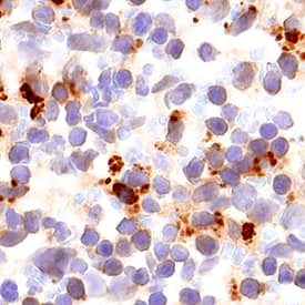
Akt2 in Mouse Spleen. Akt2 was detected in perfusion fixed frozen sections of mouse spleen using Goat Anti-Human/Mouse/Rat Akt2 Antigen Affinity-purified Polyclonal Antibody (Catalog # AF23151) at 3 µg/mL for 1 hour at room temperature followed by incubation with the Anti-Goat IgG VisUCyte™ HRP Polymer Antibody (Catalog # VC004). Tissue was stained using DAB (brown) and counterstained with hematoxylin (blue). Specific staining was localized to cytoplasm and nuclei. View our protocol for IHC Staining with VisUCyte HRP Polymer Detection Reagents.
 View Larger
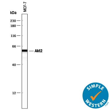
Detection of Human Akt2 by Simple WesternTM. Simple Western lane view shows lysates of MCF-7 human breast cancer cell line, loaded at 0.2 mg/mL. A specific band was detected for Akt2 at approximately 60 kDa (as indicated) using 5 µg/mL of Goat Anti-Human/Mouse/Rat Akt2 Antigen Affinity-purified Polyclonal Antibody (Catalog # AF23151) followed by 1:50 dilution of HRP-conjugated Anti-Goat IgG Secondary Antibody (Catalog # HAF109). This experiment was conducted under reducing conditions and using the 12-230 kDa separation system.
Preparation and Storage
- 12 months from date of receipt, -20 to -70 °C as supplied.
- 1 month, 2 to 8 °C under sterile conditions after reconstitution.
- 6 months, -20 to -70 °C under sterile conditions after reconstitution.
Background: Akt2
The serine/threonine kinase Akt, also known as protein kinase B (PKB), is a central regulator of such diverse cellular processes as glucose uptake, cell cycle progression, and apoptosis. In mammals, three highly homologous members define the Akt family: Akt1 (PKB alpha ), Akt2 (PKB beta ), and Akt3 (PKB gamma ). Akt2 is expressed predominantly in insulin target tissues such as liver, skeletal muscle, and fat. All three Akts contain an amino-terminal pleckstrin homology domain, a central kinase domain, and a carboxyl-terminal regulatory domain.
Product Datasheets
Citations for Human/Mouse/Rat Akt2 Antibody
R&D Systems personnel manually curate a database that contains references using R&D Systems products. The data collected includes not only links to publications in PubMed, but also provides information about sample types, species, and experimental conditions.
4
Citations: Showing 1 - 4
Filter your results:
Filter by:
-
TIM-3 Suppresses Anti-CD3/CD28-Induced TCR Activation and IL-2 Expression through the NFAT Signaling Pathway.
Authors: Tomkowicz B, Walsh E et al.
PLoS One
Species: Human
Sample Types: blood
Applications: Simple Western -
Impaired Insulin Signaling Mediated by the Small GTPase Rac1 in Skeletal Muscle of the Leptin-Deficient Obese Mouse
Authors: Chan, MP;Takenaka, N;Satoh, T;
International journal of molecular sciences
Species: Mouse
Sample Types: Tissue Homogenates
Applications: Western Blot -
Exercise effects on gamma 3-AMPK activity, phosphorylation of Akt2 and AS160, and insulin-stimulated glucose uptake in insulin-resistant rat skeletal muscle
Authors: Mark W. Pataky, Edward B. Arias, Haiyan Wang, Xiaohua Zheng, Gregory D. Cartee
Journal of Applied Physiology
-
Involvement of the protein kinase Akt2 in insulin-stimulated Rac1 activation leading to glucose uptake in mouse skeletal muscle
Authors: N Takenaka, N Araki, T Satoh
PLoS ONE, 2019-02-08;14(2):e0212219.
Species: Mouse
Sample Types: Whole Tissue
Applications: IHC-P
FAQs
No product specific FAQs exist for this product, however you may
View all Antibody FAQsReviews for Human/Mouse/Rat Akt2 Antibody
There are currently no reviews for this product. Be the first to review Human/Mouse/Rat Akt2 Antibody and earn rewards!
Have you used Human/Mouse/Rat Akt2 Antibody?
Submit a review and receive an Amazon gift card.
$25/€18/£15/$25CAN/¥75 Yuan/¥2500 Yen for a review with an image
$10/€7/£6/$10 CAD/¥70 Yuan/¥1110 Yen for a review without an image


