Human/Mouse/Rat Cyclophilin A Antibody Summary
Met1-Glu165
Accession # P62937
*Small pack size (-SP) is supplied either lyophilized or as a 0.2 µm filtered solution in PBS.
Applications
Please Note: Optimal dilutions should be determined by each laboratory for each application. General Protocols are available in the Technical Information section on our website.
Scientific Data
 View Larger
Detection of Human, Mouse, and Rat Cyclophilin A by Western Blot. Western blot shows lysates of HeLa human cervical epithelial carcinoma cell line, C2C12 mouse myoblast cell line, and Nb2-11 rat lymphoma cell line. PVDF membrane was probed with 0.1 µg/mL of Goat Anti-Human Cyclophilin A Antigen Affinity-purified Polyclonal Antibody (Catalog # AF3589) followed by HRP-conjugated Anti-Goat IgG Secondary Antibody (Catalog # HAF017). A specific band was detected for Cyclophilin A at approximately 18 kDa (as indicated). This experiment was conducted under reducing conditions and using Immunoblot Buffer Group 1.
 View Larger
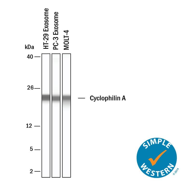
Detection of Human Cyclophilin A by Simple WesternTM. Simple Western shows lysates of Exosome Standards (HT‑29) (NBP3-11685), Exosome Standards (PC‑3) (NBP2-49856) and MOLT‑4 human acute lymphoblastic leukemia cell line, loaded at 0.5 mg/ml. A specific band was detected for Cyclophilin A at approximately 23 kDa (as indicated) using 50 µg/mL of Goat Anti-Human/Mouse/Rat Cyclophilin A Antigen Affinity-purified Polyclonal Antibody (Catalog # AF3589). This experiment was conducted under reducing conditions and using the 2-40kDa separation system.
 View Larger
Cyclophilin A in PANC-1 Human Cell Line. Cyclophilin A was detected in immersion fixed PANC-1 human pancreatic carcinoma cell line using Goat Anti-Human/Mouse/Rat Cyclophilin A Antigen Affinity-purified Polyclonal Antibody (Catalog # AF3589) at 10 µg/mL for 3 hours at room temperature. Cells were stained using the Northern-Lights™ 557-conjugated Anti-Goat IgG Secondary Antibody (red; Catalog # NL001) and counterstained with DAPI (blue). Specific staining was localized to nuclei and cytoplasm. View our protocol for Fluorescent ICC Staining of Cells on Coverslips.
 View Larger
Detection of Human and Mouse Cyclophilin A by Simple WesternTM. Simple Western lane view shows lysates of HeLa human cervical epithelial carcinoma cell line and C2C12 mouse myoblast cell line, loaded at 0.2 mg/mL. A specific band was detected for Cyclophilin A at approximately 25 kDa (as indicated) using 5 µg/mL of Goat Anti-Human/Mouse/Rat Cyclophilin A Antigen Affinity-purified Polyclonal Antibody (Catalog # AF3589) followed by 1:50 dilution of HRP-conjugated Anti-Goat IgG Secondary Antibody (Catalog # HAF109). This experiment was conducted under reducing conditions and using the 12-230 kDa separation system.
Reconstitution Calculator
Preparation and Storage
- 12 months from date of receipt, -20 to -70 °C as supplied.
- 1 month, 2 to 8 °C under sterile conditions after reconstitution.
- 6 months, -20 to -70 °C under sterile conditions after reconstitution.
Background: Cyclophilin A
Cyclophilin A, also called Peptidyl-prolyl Isomerase A, PPIA, CYPA, and CYPH, was originally characterized for its ability to catalyze the transition between cis- and trans- proline residues critical for proper folding of proteins (1). Cyclophilin is also incorporated into many viruses, including HIV-1, where it has been speculated to be involved in functions such as viral assembly and infectivity (2). The immunosuppressive activity of cyclosporins has been correlated with their ability to form complexes with cyclophilins that inhibit calcineurin phosphatase activity (3) and prevent incorporation of cyclophilin into viral particles (4). The cyclosporin/cyclophilin complex selectively binds and inactivates calcineurin (3, 5), making it a useful inhibitor for studying calcineurin activity.
- Hamilton, G.S. and J.P. Steiner (1998) J. Med. Chem. 41:5119.
- Cantin, R. et al. (2005) J. Virology 79:6577.
- Liu, J. et al. (1992) Biochemistry 31:3896.
- Wiegers K. and H.G. Krausslich (2002) Virology 294:289.
- Liu, J. et al. (1991) Cell 66:807.
Product Datasheets
Citations for Human/Mouse/Rat Cyclophilin A Antibody
R&D Systems personnel manually curate a database that contains references using R&D Systems products. The data collected includes not only links to publications in PubMed, but also provides information about sample types, species, and experimental conditions.
2
Citations: Showing 1 - 2
Filter your results:
Filter by:
-
Human Parvovirus Infection of Human Airway Epithelia Induces Pyroptotic Cell Death by Inhibiting Apoptosis
Authors: Xuefeng Deng, Wei Zou, Min Xiong, Zekun Wang, John F. Engelhardt, Shui Qing Ye et al.
Journal of Virology
-
The Novel Extracellular Cyclophilin A (CyPA) - Inhibitor MM284 Reduces Myocardial Inflammation and Remodeling in a Mouse Model of Troponin I -Induced Myocarditis.
Authors: Heinzmann D, Bangert A, Muller A, von Ungern-Sternberg S, Emschermann F, Schonberger T, Chatterjee M, Mack A, Klingel K, Kandolf R, Malesevic M, Borst O, Gawaz M, Langer H, Katus H, Fischer G, May A, Kaya Z, Seizer P
PLoS ONE, 2015-04-20;10(4):e0124606.
Species: Mouse
Sample Types: Whole Tissue
Applications: IHC
FAQs
No product specific FAQs exist for this product, however you may
View all Antibody FAQsReviews for Human/Mouse/Rat Cyclophilin A Antibody
There are currently no reviews for this product. Be the first to review Human/Mouse/Rat Cyclophilin A Antibody and earn rewards!
Have you used Human/Mouse/Rat Cyclophilin A Antibody?
Submit a review and receive an Amazon gift card.
$25/€18/£15/$25CAN/¥75 Yuan/¥2500 Yen for a review with an image
$10/€7/£6/$10 CAD/¥70 Yuan/¥1110 Yen for a review without an image

