Human/Mouse/Rat Fetuin B Antibody Summary
Met19-Pro382
Accession # Q9UGM5
*Small pack size (-SP) is supplied either lyophilized or as a 0.2 µm filtered solution in PBS.
Applications
Please Note: Optimal dilutions should be determined by each laboratory for each application. General Protocols are available in the Technical Information section on our website.
Scientific Data
 View Larger
Detection of Human, Mouse, and Rat Fetuin B by Western Blot. Western blot shows human serum, human placenta tissue lysate, mouse serum, and rat placenta tissue lysate. PVDF membrane was probed with 0.25 µg/mL of Goat Anti-Human/Mouse/Rat Fetuin B Antigen Affinity-purified Polyclonal Antibody (Catalog # AF1725) followed by HRP-conjugated Anti-Goat IgG Secondary Antibody (HAF017). A specific band was detected for Fetuin B at approximately 55 kDa (as indicated). This experiment was conducted under reducing conditions and using Immunoblot Buffer Group 1.
 View Larger
Detection of Human Fetuin B by Simple WesternTM. Simple Western lane view shows lysates of human placenta tissue and rat placenta tissue, loaded at 0.2 mg/mL. A specific band was detected for Fetuin B at approximately 64 kDa (as indicated) using 2.5 µg/mL of Goat Anti-Human/Mouse/Rat Fetuin B Antigen Affinity-purified Polyclonal Antibody (Catalog # AF1725) followed by 1:50 dilution of HRP-conjugated Anti-Goat IgG Secondary Antibody (HAF109). This experiment was conducted under reducing conditions and using the 12-230 kDa separation system.
 View Larger
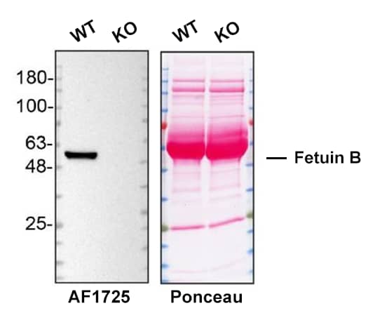
Western Blot Shows Mouse Fetuin B Specificity Using Knockout Cell Line. Western blot shows WT and Fetuin B KO serum from mouse. Nitrocellulose membrane was probed with 0.2 µg/mL of Goat Anti-Human/Mouse/Rat Fetuin B Antigen Affinity-purified Polyclonal Antibody (Catalog # AF1725) followed by HRP-conjugated donkey anti-goat IgG Secondary Antibody. A specific band was detected for Fetuin B at approximately 60 kDa (as indicated), but is not detectable in knockout sera. The Ponceau stained transfer of the blot is shown. This experiment was conducted under reducing conditions. Image, protocol, and testing courtesy of YCharOS Inc. See ycharos.com for additional details.
Reconstitution Calculator
Preparation and Storage
- 12 months from date of receipt, -20 to -70 °C as supplied.
- 1 month, 2 to 8 °C under sterile conditions after reconstitution.
- 6 months, -20 to -70 °C under sterile conditions after reconstitution.
Background: Fetuin B
Fetuins are members of the cystatin superfamily of cysteine protease inhibitors (1‑3). Additional members of this superfamily are kininogen and histidine-rich glycoprotein. Fetuin A and B are two known members of the fetuin family. Hepatocytes are believed to be the principal cellular source, but other cell types also express it (4, 5). Fetuin A, also known as alpha 2-Heremans-Schmid glycoprotein, is an inhibitor of basic calcium phosphate precipitation and a negative acute-phase protein (6, 7). Normal circulating levels of Fetuin A in adults (300‑600 ug/mL) fall significantly (30‑50%) during injury and infection (7). Fetuin B is a newer member whose function is not fully characterized (1, 2). Fetuin A and B display similarities and differences in their characteristics. Fetuin B exhibits reduction of calcification, while both mRNA levels were down‑regulated during the acute phase in inflammation-induced rats (4). However, they share only 20% amino acid sequence identity (2). The amounts of Fetuin B in human serum, unlike Fetuin A, vary with gender and are higher in females than in males (4).
- Oliver, E. et al. (1999) Genomics. 57:352.
- Oliver, E. et al. (2000) Biochem. J. 350:589.
- Kellemann, J. et al. 1989, J. Biol. Chem. 264:14121.
- Denecke, B. et al. (2003) Biochem. J. 376:135.
- Schäfer, C. et al. (2003) J. Clin. Invest. 112:357.
- Dziegielewska, K. M. et al. (1996) Histochem. Cell Biol. 106:319.
- Gangneux, C. et al. (2003) Nucleic Acids Res. 31:5957.
Product Datasheets
Citation for Human/Mouse/Rat Fetuin B Antibody
R&D Systems personnel manually curate a database that contains references using R&D Systems products. The data collected includes not only links to publications in PubMed, but also provides information about sample types, species, and experimental conditions.
1 Citation: Showing 1 - 1
-
The liver of woodchucks chronically infected with the woodchuck hepatitis virus contains foci of virus core antigen-negative hepatocytes with both altered and normal morphology.
Authors: Xu C, Yamamoto T, Zhou T, Aldrich CE, Frank K, Cullen JM, Jilbert AR, Mason WS
Virology, 2006-10-31;359(2):283-94.
Species: Human, Woodchuck
Sample Types: Tissue Homogenates, Whole Cells, Whole Tissue
Applications: ICC, IHC-P, Western Blot
FAQs
No product specific FAQs exist for this product, however you may
View all Antibody FAQsReviews for Human/Mouse/Rat Fetuin B Antibody
There are currently no reviews for this product. Be the first to review Human/Mouse/Rat Fetuin B Antibody and earn rewards!
Have you used Human/Mouse/Rat Fetuin B Antibody?
Submit a review and receive an Amazon gift card.
$25/€18/£15/$25CAN/¥75 Yuan/¥2500 Yen for a review with an image
$10/€7/£6/$10 CAD/¥70 Yuan/¥1110 Yen for a review without an image

