Human/Mouse/Rat HHEX Antibody Summary
Thr111-Gly270
Accession # Q03014
Applications
Please Note: Optimal dilutions should be determined by each laboratory for each application. General Protocols are available in the Technical Information section on our website.
Scientific Data
 View Larger
Detection of Human, Mouse, and Rat HHEX by Western Blot. Western blot shows lysates of HepG2 human hepatocellular carcinoma cell line, BaF3 mouse pro-B cell line, and H4-II-E-C3 rat hepatoma cell line. PVDF membrane was probed with 1 µg/mL of Rabbit Anti-Human/Mouse/Rat HHEX Monoclonal Antibody (Catalog # MAB83771) followed by HRP-conjugated Anti-Rabbit IgG Secondary Antibody (Catalog # HAF008). A specific band was detected for HHEX at approximately 37 kDa (as indicated). This experiment was conducted under reducing conditions and using Immunoblot Buffer Group 1.
 View Larger
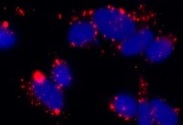
HHEX in K562 Human Cell Line. HHEX was detected in immersion fixed K562 human chronic myelogenous leukemia cell line using Rabbit Anti-Human/Mouse/Rat HHEX Polyclonal Antibody (Catalog # MAB83771) at 3 µg/mL for 3 hours at room temperature. Cells were stained using the NorthernLights™ 557-conjugated Anti-Rabbit IgG Secondary Antibody (red; Catalog # NL004) and counterstained with DAPI (blue). Specific staining was localized to nuclei. View our protocol for Fluorescent ICC Staining of Non-adherent Cells.
 View Larger
Detection of HHEX in A549 Human Cell Line by Flow Cytometry. A549 human lung carcinoma cell line was stained with Rabbit Anti-Human/Mouse/Rat HHEX Monoclonal Antibody (Catalog # MAB83771, filled histogram) or isotype control antibody (Catalog # AB-105-C, open histogram), followed by Phycoerythrin-conjugated Anti-Rabbit IgG Secondary Antibody (Catalog # F0110). To facilitate intracellular staining, cells were fixed and permeabilized with FlowX FoxP3 Fixation & Permeabilization Buffer Kit (Catalog # FC012). View our protocol for Staining Intracellular Molecules.
 View Larger
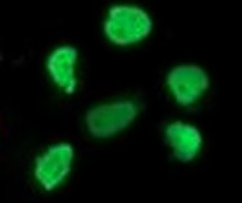
HHEX in iBJ6 iPS Cell Line. HHEX was detected in immersion fixed iBJ6 iPS cell line differentiated into hepatocytes using Rabbit Anti-Human/Mouse/Rat HHEX Polyclonal Antibody (Catalog # MAB83771) at 2 µg/mL for 3 hours at room temperature. Cells were stained using the NorthernLights™ 557-conjugated Anti-Rabbit IgG Secondary Antibody (red; Catalog # NL004) and counterstained with DAPI (blue). Specific staining was localized to nuclei. View our protocol for Fluorescent ICC Staining of Cells on Coverslips.
Reconstitution Calculator
Preparation and Storage
- 12 months from date of receipt, -20 to -70 °C as supplied.
- 1 month, 2 to 8 °C under sterile conditions after reconstitution.
- 6 months, -20 to -70 °C under sterile conditions after reconstitution.
Background: HHEX
Hematopoietically-expressed homeobox protein (HHEX), also known as HEX, PRH and PRHX, is a 35-40 kDa member of the Homeobox family of transcription factors. Family members are distinguished by an evolutionarily conserved DNA-binding homeodomain of 60 amino acids (aa), which for HHEX spans aa 137-196. Human HHEX was initially isolated from hematopoietic tissue, and is present in several hematopoietic progenitors, where its expression is down-regulated during terminal cell differentiation. HHEX is also expressed in the anterior visceral endoderm during early mouse development, and in some adult tissues of endodermal origin, including liver, lung and thyroid. HHEX knockout in mice is embryonic lethal, with impaired forebrain, liver and thyroid development. Human HHEX is 270 aa in length, and over aa 111-270, shares 93% and 95% identity with mouse and rat HHEX, respectively.
Product Datasheets
Citations for Human/Mouse/Rat HHEX Antibody
R&D Systems personnel manually curate a database that contains references using R&D Systems products. The data collected includes not only links to publications in PubMed, but also provides information about sample types, species, and experimental conditions.
6
Citations: Showing 1 - 6
Filter your results:
Filter by:
-
Elucidation of HHEX in pancreatic endoderm differentiation using a human iPSC differentiation model
Authors: Ito, R;Kimura, A;Hirose, Y;Hatano, Y;Mima, A;Mae, SI;Keidai, Y;Nakamura, T;Fujikura, J;Nishi, Y;Ohta, A;Toyoda, T;Inagaki, N;Osafune, K;
Scientific reports
Species: Human
Sample Types: Cell Lysates
Applications: Western Blot -
Expansion of ventral foregut is linked to changes in the enhancer landscape for organ-specific differentiation
Authors: YF Wong, Y Kumar, M Proks, JAR Herrera, MM Rothová, RS Monteiro, S Pozzi, RE Jennings, NA Hanley, WA Bickmore, JM Brickman
Nature Cell Biology, 2023-01-23;0(0):.
Species: Human
Sample Types: Whole Cells
Applications: ChIP -
CRISPR screening uncovers a central requirement for HHEX in pancreatic lineage commitment and plasticity restriction
Authors: D Yang, H Cho, Z Tayyebi, A Shukla, R Luo, G Dixon, V Ursu, S Stransky, DM Tremmel, SD Sackett, R Koche, SJ Kaplan, QV Li, J Park, Z Zhu, BP Rosen, J Pulecio, ZD Shi, Y Bram, RE Schwartz, JS Odorico, S Sidoli, CV Wright, CS Leslie, D Huangfu
Nature Cell Biology, 2022-07-04;0(0):.
Species: Human
Sample Types: Cell Lysates
Applications: Western Blot -
Cell-fate transition and determination analysis of mouse male germ cells throughout development
Authors: J Zhao, P Lu, C Wan, Y Huang, M Cui, X Yang, Y Hu, Y Zheng, J Dong, M Wang, S Zhang, Z Liu, S Bian, X Wang, R Wang, S Ren, D Wang, Z Yao, G Chang, F Tang, XY Zhao
Nature Communications, 2021-11-25;12(1):6839.
Species: Mouse
Sample Types: Cell Lysates
Applications: Western Blot -
Hhex Directly Represses BIM-Dependent Apoptosis to Promote NK Cell Development and Maintenance
Authors: W Goh, S Scheer, JT Jackson, S Hediyeh-Za, RB Delconte, IS Schuster, CE Andoniou, J Rautela, MA Degli-Espo, MJ Davis, MP McCormack, SL Nutt, ND Huntington
Cell Rep, 2020-10-20;33(3):108285.
Species: Mouse
Sample Types: Cell Lysates
Applications: Western Blot -
Single-Cell RNA-Sequencing-Based CRISPRi Screening Resolves Molecular Drivers of Early Human Endoderm Development
Authors: RMJ Genga, EM Kernfeld, KM Parsi, TJ Parsons, MJ Ziller, R Maehr
Cell Rep, 2019-04-16;27(3):708-718.e10.
Species: Human
Sample Types: Whole Cells
Applications: ICC
FAQs
No product specific FAQs exist for this product, however you may
View all Antibody FAQsReviews for Human/Mouse/Rat HHEX Antibody
Average Rating: 5 (Based on 2 Reviews)
Have you used Human/Mouse/Rat HHEX Antibody?
Submit a review and receive an Amazon gift card.
$25/€18/£15/$25CAN/¥75 Yuan/¥2500 Yen for a review with an image
$10/€7/£6/$10 CAD/¥70 Yuan/¥1110 Yen for a review without an image
Filter by:


