Human/Mouse/Rat HTRA2/Omi Antibody Summary
Ala134-Glu458
Accession # O43464
Applications
Please Note: Optimal dilutions should be determined by each laboratory for each application. General Protocols are available in the Technical Information section on our website.
Scientific Data
 View Larger
Detection of Human/Mouse/Rat HTRA2/Omi by Western Blot. Western blot shows lysates of PC-12 rat adrenal pheochromocytoma cell line, Jurkat human acute T cell leukemia cell line, HeLa human cervical epithelial carcinoma cell line, L-929 mouse fibroblast cell line, and C2C12 mouse myoblast cell line. PVDF membrane was probed with 0.25 µg/mL of Rabbit Anti-Human/Mouse/Rat HTRA2/Omi Antigen Affinity-purified Polyclonal Antibody (Catalog # AF1458) followed by HRP-conjugated Anti-Rabbit IgG Secondary Antibody (Catalog # HAF008). Specific bands were detected for HTRA2/Omi at approximately 36 and 49 kDa (as indicated). This experiment was conducted under reducing conditions and using Immunoblot Buffer Group 2.
 View Larger
HTRA2/Omi in Jurkat Human Cell Line. HTRA2/Omi was detected in immersion fixed Jurkat human acute T cell leukemia cell line stimulated with staurosporin using Rabbit Anti-Human/Mouse/Rat HTRA2/Omi Antigen Affinity-purified Polyclonal Antibody (Catalog # AF1458) at 10 µg/mL for 3 hours at room temperature. Cells were stained using the NorthernLights™ 557-conjugated Anti-Rabbit IgG Secondary Antibody (yellow; Catalog # NL004) and counterstained with DAPI (blue). View our protocol for Fluorescent ICC Staining of Cells on Coverslips.
 View Larger
Detection of Human and Mouse HTRA2/Omi by Simple WesternTM. Simple Western lane view shows lysates of C2C12 mouse myoblast cell line and HeLa human cervical epithelial carcinoma cell line, loaded at 0.2 mg/mL. Specific bands were detected for HTRA2/Omi at approximately 51 kDa (precursor) and 41 kDa (processed) (as indicated) using 2.5 µg/mL of Rabbit Anti-Human/Mouse/Rat HTRA2/Omi Antigen Affinity-purified Polyclonal Antibody (Catalog # AF1458). This experiment was conducted under reducing conditions and using the 12-230 kDa separation system.
 View Larger
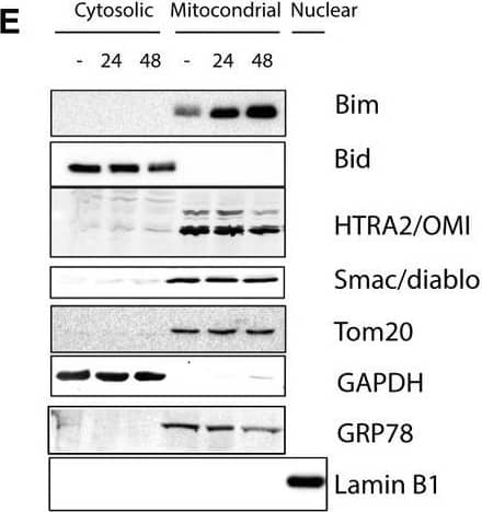
Detection of Human HTRA2/Omi by Western Blot Caspases are not activated by FGFR inhibition in FGFR2‐mutant EC cells. (A) Western blots showing total caspase‐3 and caspase‐7 in response to treatment with 300 nm BGJ398 for up to 72 h. Tubulin serves as a loading control. *Denotes nonspecific band. (B) AN3CA and JHUEM2 cells were pretreated with 100 μm Z‐VAD‐FMK for 1 h prior to the addition of DMSO, 1 μm PD173074 (PD), 300 nm BGJ398 (BGJ) or 300 nm AZD4547 (AZD) for 72 h. Cell death was detected by staining cells with Annexin V. The mean percentage of Annexin V‐positive cells from three independent experiments (each performed in triplicate) is shown along with SD. (C) Western blot showing cleavage of caspase‐3 in AN3CA and JHUEM2 cells treated with 1 μm actinomycin D (Act D) or staurosporine (STS), respectively, for 24 h. (D) Mean percentage of AN3CA and JHUEM2 cells showing Annexin V‐positive staining following treatment with 100 μm Z‐VAD‐FMK alone or 1 h prior to treatment with 1 μm Act D and STS for 24 h. The mean from three independent experiments (each performed in triplicate) is shown along with SD. (E) Western blot showing staining of Bim, Bid, HTRA2/OMI and Smac/diablo in cytosolic and mitochondrial fractions of JHUEM2 cells treated with 300 nm BGJ398 for 24 and 48 h. Tom20 serves as a marker of the mitochondrial fraction, GRP78 as a marker of the ER, Lamin B1 as a marker of the nuclear fraction and GAPDH as a marker of the cytosolic fraction. (F) Western blot showing staining of AIF in mitochondrial and nuclear fractions of AN3CA cells treated with 1 μm PD173074 for 48 h. Cox IV and PARP serve as mitochondrial and nuclear markers, respectively. Image collected and cropped by CiteAb from the following open publication (https://pubmed.ncbi.nlm.nih.gov/30537101), licensed under a CC-BY license. Not internally tested by R&D Systems.
 View Larger
Detection of Mouse HTRA2/Omi by Western Blot Neural deletion of Htra2 is sufficient to generate neurological phenotypes.(A) Exons 2 to 4 of Htra2 were flanked with loxP sites, with a FRT flanked neo cassette 3′ to exon 4. Expression of FlpE causes deletion of the selection cassette. Cre-mediated deletion causes excision of exons 2 to 4. Small arrows beneath the allele constructs denote the position of genotyping primers. (B) PCR from genomic DNA can distinguish WT (+, arrow, 279 bp), KO (–, filled arrowhead, 358 bp) and floxed (f, empty arrowhead, 313 bp) alleles of Htra2. (C) Western blot analysis confirmed loss of HTRA2 protein (arrow) in all tissues of HTRA2 KO mice and reduction in brain of NesKO mice (arrowheads denote non-specific bands). The levels of HTRA2 protein in NesKO spleen and thymus were comparable with NesWT. Cx: cortex, Mb: midbrain, Hb: hindbrain. PHB2 was used as a loading control. (D) HTRA2 KO mice and NesKO mice were smaller than WT littermates by comparison. The size of the thymus and spleen was reduced although brain was relatively normal in size (representative animals shown at P30, scale bar: 1 cm.). (E) Body weight of HTRA2 KO and NesKO mice did not increase beyond P18 (n = 56 (HTRA2 WT), 62 (HTRA2 KO), 35 (NesWT), 25 (NesKO), error bars indicate SEM). Image collected and cropped by CiteAb from the following open publication (https://pubmed.ncbi.nlm.nih.gov/25531304), licensed under a CC-BY license. Not internally tested by R&D Systems.
Reconstitution Calculator
Preparation and Storage
- 12 months from date of receipt, -20 to -70 °C as supplied.
- 1 month, 2 to 8 °C under sterile conditions after reconstitution.
- 6 months, -20 to -70 °C under sterile conditions after reconstitution.
Background: HTRA2/Omi
HtrA2/Omi is the mammalian homologue of bacterial high temperature requirement protein (HtrA). HtrA2/Omi localizes to the mitochondria and is processed to expose an amino-terminal Reaper-like motif similar to SMAC/Diablo. HtrA2/Omi is released from the mitochondria in response to apoptotic insult and can interact with the BIR2 or BIR3 domains of XIAP to relieve caspase-IAP inhibition. This effect can be measured by reversing XIAP-BIR2 (R&D Systems, Catalog # 786-XB) inhibition of Caspase-7 (R&D Systems, Catalog # 823-C7) cleavage of a fluorogenic peptide (DEVD-AFC, MP Bio, Catalog # AFC-138). IC50 values for this effect are typically between 0.2 and 1.5 μM. HtrA2/Omi is trimeric and functions as a serine protease. The serine protease activity may play a more central role in apoptosis than its IAP antagonizing function. A PDZ domain regulates the serine protease activity by blocking access to the active site. The specificity of the protease is yet to be defined and no endogenous substrates are known to date.
- Suzuki, Y. et al. (2001) Mol. Cell. 8:613.
- van Loo, G. et al. (2002) Cell Death & Diff. 9:20.
- Hedge, R. et al. (2001) J. Biol. Chem. 277:432.
- Verhagen, A. et al. (2001) J. Biol. Chem. 277:445.
- Martins, L. et al. (2002) J. Biol. Chem. 277:439.
- Silke, J., and A. Verhagen (2002) Cell Death & Diff. 9:362.
- Savopoulos, J. et al. (2000) Protein Expression & Purification 19:227.
Product Datasheets
Citations for Human/Mouse/Rat HTRA2/Omi Antibody
R&D Systems personnel manually curate a database that contains references using R&D Systems products. The data collected includes not only links to publications in PubMed, but also provides information about sample types, species, and experimental conditions.
33
Citations: Showing 1 - 10
Filter your results:
Filter by:
-
Protease-independent control of parthanatos by HtrA2/Omi
Authors: Jonas Wei beta, Michelle Heib, Thiemo Korn, Justus Hoyer, Johaiber Fuchslocher Chico, Susann Voigt et al.
Cellular and Molecular Life Sciences
-
Subcellular origin of mitochondrial DNA deletions in human skeletal muscle
Authors: Amy E. Vincent, Hannah S. Rosa, Kamil Pabis, Conor Lawless, Chun Chen, Anne Grünewald et al.
Annals of Neurology
-
The protease Omi regulates mitochondrial biogenesis through the GSK3B/PGC-1a pathway
Authors: Xu R, Hu Q, Ma Q et al.
Cell Death Dis
-
Increased Active OMI/HTRA2 Serine Protease Displays a Positive Correlation with Cholinergic Alterations in the Alzheimer’s Disease Brain
Authors: Taher Darreh-Shori, Sareh Rezaeianyazdi, Erica Lana, Sumonto Mitra, Anna Gellerbring, Azadeh Karami et al.
Molecular Neurobiology
-
The multi-subunit GID/CTLH E3 ubiquitin ligase promotes cell proliferation and targets the transcription factor Hbp1 for degradation
Authors: F Lampert, D Stafa, A Goga, MV Soste, S Gilberto, N Olieric, P Picotti, M Stoffel, M Peter
Elife, 2018-06-18;7(0):.
-
Parkinson’s disease-associated mutations in DJ-1 modulate its dimerization in living cells
Authors: Mariaelena Repici, Kornelis R. Straatman, Nadia Balduccio, Francisco J. Enguita, Tiago F. Outeiro, Flaviano Giorgini
Journal of Molecular Medicine
-
Bcl‐2 inhibitors enhance FGFR inhibitor‐induced mitochondrial‐dependent cell death in FGFR2‐mutant endometrial cancer
Authors: Leisl M. Packer, Samantha J. Stehbens, Vanessa F. Bonazzi, Jennifer H. Gunter, Robert J. Ju, Micheal Ward et al.
Molecular Oncology
-
Diastolic dysfunction in Alzheimer's disease model mice is associated with A?-amyloid aggregate formation and mitochondrial dysfunction
Authors: Aishwarya, R;Abdullah, CS;Remex, NS;Bhuiyan, MAN;Lu, XH;Dhanesha, N;Stokes, KY;Orr, AW;Kevil, CG;Bhuiyan, MS;
Scientific reports
Species: Mouse
Sample Types: Cell Lysates
Applications: Western Blot -
Pleiotropic effects of mdivi-1 in altering mitochondrial dynamics, respiration, and autophagy in cardiomyocytes
Authors: R Aishwarya, S Alam, CS Abdullah, M Morshed, SS Nitu, M Panchatcha, S Miriyala, CG Kevil, MS Bhuiyan
Redox Biol, 2020-07-26;36(0):101660.
Species: Rat
Sample Types: Cell Lysates, Protein
Applications: Western Blot -
Cytosolic Trapping of a Mitochondrial Heat Shock Protein Is an Early Pathological Event in Synucleinopathies
Authors: ÉM Szeg?, A Dominguez-, E Gerhardt, A König, DJ Koss, W Li, R Pinho, C Fahlbusch, M Johnson, P Santos, A Villar-Piq, T Thom, S Rizzoli, M Schmitz, J Li, I Zerr, J Attems, O Jahn, TF Outeiro
Cell Rep, 2019-07-02;28(1):65-77.e6.
Species: Rat
Sample Types: Cell Lysates
Applications: Western Blot -
PARL mediates Smac proteolytic maturation in mitochondria to promote apoptosis
Authors: S Saita, H Nolte, KU Fiedler, H Kashkar, AS Venne, RP Zahedi, M Krüger, T Langer
Nat. Cell Biol, 2017-03-13;0(0):.
Species: Human
Sample Types: Cell Lysates
Applications: Western Blot -
Protease Omi cleaving Hax-1 protein contributes to OGD/R-induced mitochondrial damage in neuroblastoma N2a cells and cerebral injury in MCAO mice.
Authors: Wu J, Li M, Cao L, Sun M, Chen D, Ren H, Xia Q, Tao Z, Qin Z, Hu Q, Wang G
Acta Pharmacol Sin, 2015-08-24;36(9):1043-52.
Species: Mouse
Sample Types: Tissue Homogenates
Applications: Western Blot -
Protease Omi facilitates neurite outgrowth in mouse neuroblastoma N2a cells by cleaving transcription factor E2F1.
Authors: Ma Q, Hu Q, Xu R, Zhen X, Wang G
Acta Pharmacol Sin, 2015-08-01;36(8):966-75.
Species: Human, Mouse
Sample Types: Cell Lysates, Tissue Homogenates
Applications: Western Blot -
Heat shock inhibition of CDK5 increases NOXA levels through miR-23a repression.
Authors: Morey T, Roufayel R, Johnston D, Fletcher A, Mosser D
J Biol Chem, 2015-03-31;290(18):11443-54.
Species: Human
Sample Types: Cell Lysates
Applications: Western Blot -
Neural-specific deletion of Htra2 causes cerebellar neurodegeneration and defective processing of mitochondrial OPA1.
Authors: Patterson, Victoria, Zullo, Alfred J, Koenig, Claire, Stoessel, Sean, Jo, Hakryul, Liu, Xinran, Han, Jinah, Choi, Murim, DeWan, Andrew T, Thomas, Jean-Leo, Kuan, Chia-Yi, Hoh, Josephin
PLoS ONE, 2014-12-22;9(12):e115789.
Species: Mouse
Sample Types: Cell Lysates
Applications: Western Blot -
PINK1 kinase catalytic activity is regulated by phosphorylation on serines 228 and 402.
Authors: Aerts L, Craessaerts K, de Strooper B, Morais V
J Biol Chem, 2014-12-19;290(5):2798-811.
Species: Human
Sample Types: Cell Lysates
Applications: Western Blot -
Influenza A virus protein PB1-F2 translocates into mitochondria via Tom40 channels and impairs innate immunity.
Authors: Yoshizumi T, Ichinohe T, Sasaki O, Otera H, Kawabata S, Mihara K, Koshiba T
Nat Commun, 2014-08-20;5(0):4713.
Species: Human
Sample Types: Cell Lysates
Applications: Western Blot -
The ubiquitin-conjugating enzymes UBE2N, UBE2L3 and UBE2D2/3 are essential for Parkin-dependent mitophagy.
Authors: Geisler S, Vollmer S, Golombek S, Kahle P
J Cell Sci, 2014-06-06;127(0):3280-93.
Species: Human
Sample Types: Whole Cells
Applications: ICC -
p53-mediated activation of the mitochondrial protease HtrA2/Omi prevents cell invasion.
Authors: Yamauchi S, Hou Y, Guo A, Hirata H, Nakajima W, Yip A, Yu C, Harada I, Chiam K, Sawada Y, Tanaka N, Kawauchi K
J Cell Biol, 2014-03-24;204(7):1191-207.
Species: Mouse
Sample Types: Whole Cells
Applications: IHC -
The accumulation of misfolded proteins in the mitochondrial matrix is sensed by PINK1 to induce PARK2/Parkin-mediated mitophagy of polarized mitochondria.
Authors: Jin, Seok Min, Youle, Richard
Autophagy, 2013-09-05;9(11):1750-7.
Species: Human
Sample Types: Cell Lysates
Applications: Western Blot -
PINK1 is degraded through the N-end rule pathway.
Authors: Yamano, Koji, Youle, Richard
Autophagy, 2013-04-17;9(11):1758-69.
Species: Human
Sample Types: Cell Lysates
Applications: Western Blot -
PINK1/Parkin-mediated mitophagy is dependent on VDAC1 and p62/SQSTM1.
Authors: Geisler S, Holmstrom KM, Skujat D, Fiesel FC, Rothfuss OC, Kahle PJ, Springer W
Nat. Cell Biol., 2010-01-24;12(2):119-31.
Species: Human
Sample Types: Whole Cells
Applications: ICC -
Novel mitochondrial substrates of omi indicate a new regulatory role in neurodegenerative disorders.
Authors: Johnson F, Kaplitt MG
PLoS ONE, 2009-09-18;4(9):e7100.
Species: Human
Sample Types: Cell Lysates
Applications: Western Blot -
Enhanced HtrA2/Omi expression in oxidative injury to retinal pigment epithelial cells and murine models of neurodegeneration.
Authors: Ding X, Patel M, Shen D, Herzlich AA, Cao X, Villasmil R, Klupsch K, Tuo J, Downward J, Chan CC
Invest. Ophthalmol. Vis. Sci., 2009-05-14;50(10):4957-66.
Species: Human, Mouse
Sample Types: Whole Cells, Whole Tissue
Applications: ICC, IHC-Fr -
Identification of a novel protein MICS1 that is involved in maintenance of mitochondrial morphology and apoptotic release of cytochrome c.
Authors: Oka T, Sayano T, Tamai S, Yokota S, Kato H, Fujii G, Mihara K
Mol. Biol. Cell, 2008-04-16;19(6):2597-608.
Species: Human
Sample Types: Whole Cells
Applications: ICC -
HtrA2 regulates beta-amyloid precursor protein (APP) metabolism through endoplasmic reticulum-associated degradation.
Authors: Huttunen HJ, Guenette SY, Peach C, Greco C, Xia W, Kim DY, Barren C, Tanzi RE, Kovacs DM
J. Biol. Chem., 2007-08-06;282(38):28285-95.
Species: Hamster
Sample Types: Cell Lysates, Whole Cells
Applications: ICC, Western Blot -
Yersinia YopP-induced apoptotic cell death in murine dendritic cells is partially independent from action of caspases and exhibits necrosis-like features.
Authors: Grobner S, Autenrieth SE, Soldanova I, Gunst DS, Schaller M, Bohn E, Muller S, Leverkus M, Wesselborg S, Autenrieth IB, Borgmann S
Apoptosis, 2006-11-01;11(11):1959-68.
Species: Mouse
Sample Types: Cell Culture Supernates
Applications: Western Blot -
Induction of BIM(EL) following growth factor withdrawal is a key event in caspase-dependent apoptosis of 661W photoreceptor cells.
Authors: Gomez-Vicente V, Doonan F, Donovan M, Cotter TG
Eur. J. Neurosci., 2006-08-01;24(4):981-90.
Species: Mouse
Sample Types: Whole Cells
Applications: ICC -
Role and regulation of nodal/activin receptor-like kinase 7 signaling pathway in the control of ovarian follicular atresia.
Authors: Wang H, Jiang JY, Zhu C, Peng C, Tsang BK
Mol. Endocrinol., 2006-05-18;20(10):2469-82.
Species: Rat
Sample Types: Cell Lysates
Applications: Western Blot -
Roscovitine-induced up-regulation of p53AIP1 protein precedes the onset of apoptosis in human MCF-7 breast cancer cells.
Authors: Wesierska-Gadek J, Gueorguieva M, Horky M
Mol. Cancer Ther., 2005-01-01;4(1):113-24.
Species: Human
Sample Types: Whole Cells
Applications: ICC -
Motoneuron resistance to apoptotic cell death in vivo correlates with the ratio between X-linked inhibitor of apoptosis proteins (XIAPs) and its inhibitor, XIAP-associated factor 1.
Authors: Perrelet D, Perrin FE, Liston P, Korneluk RG, MacKenzie A, Ferrer-Alcon M, Kato AC
J. Neurosci., 2004-04-14;24(15):3777-85.
Species: Rat
Sample Types: Tissue Homogenates
Applications: Western Blot -
The Role of Parl and HtrA2 in Striatal Neuronal Injury After Transient Global Cerebral Ischemia
Authors: Hideyuki Yoshioka, Masataka Katsu, Hiroyuki Sakata, Nobuya Okami, Takuma Wakai, Hiroyuki Kinouchi et al.
Journal of Cerebral Blood Flow & Metabolism
-
Decreased apoptosome activity with neuronal differentiation sets the threshold for strict IAP regulation of apoptosis
Authors: Kevin M. Wright, Michael W. Linhoff, Patrick Ryan Potts, Mohanish Deshmukh
The Journal of Cell Biology
FAQs
No product specific FAQs exist for this product, however you may
View all Antibody FAQsReviews for Human/Mouse/Rat HTRA2/Omi Antibody
There are currently no reviews for this product. Be the first to review Human/Mouse/Rat HTRA2/Omi Antibody and earn rewards!
Have you used Human/Mouse/Rat HTRA2/Omi Antibody?
Submit a review and receive an Amazon gift card.
$25/€18/£15/$25CAN/¥75 Yuan/¥2500 Yen for a review with an image
$10/€7/£6/$10 CAD/¥70 Yuan/¥1110 Yen for a review without an image


