Human/Mouse/Rat RelA/NF kappa B p65 Antibody Summary
Asn456-Ser551
Accession # Q04206
Applications
Please Note: Optimal dilutions should be determined by each laboratory for each application. General Protocols are available in the Technical Information section on our website.
Scientific Data
 View Larger
Detection of Human RelA/NF kappa B p65 by Western Blot. Western blot shows lysates of K562 human chronic myelogenous leukemia cell line, Daudi human Burkitt's lymphoma cell line, and LNCaP human prostate cancer cell line. PVDF membrane was probed with 1 µg/mL of Sheep Anti-Human/Mouse/Rat RelA/NF kappa B p65 Antigen Affinity-purified Polyclonal Antibody (Catalog # AF5078) followed by HRP-conjugated Anti-Sheep IgG Secondary Antibody (Catalog # HAF016). A specific band was detected for RelA/NF kappa B p65 at approximately 70 kDa (as indicated). This experiment was conducted under reducing conditions and using Immunoblot Buffer Group 1.
 View Larger
Detection of Human and Mouse RelA/NF kappa B p65 by Western Blot. Western blot shows lysates of K562 human chronic myelogenous leukemia cell line, HeLa human cervical epithelial carcinoma cell line, and Neuro-2A mouse neuroblastoma cell line. PVDF membrane was probed with 1 µg/mL of Sheep Anti-Human/Mouse/Rat RelA/NF kappa B p65 Antigen Affinity-purified Polyclonal Antibody (Catalog # AF5078) followed by HRP-conjugated Anti-Sheep IgG Secondary Antibody (Catalog # HAF016). A specific band was detected for RelA/NF kappa B p65 at approximately 70 kDa (as indicated). This experiment was conducted under reducing conditions and using Immunoblot Buffer Group 1.
 View Larger
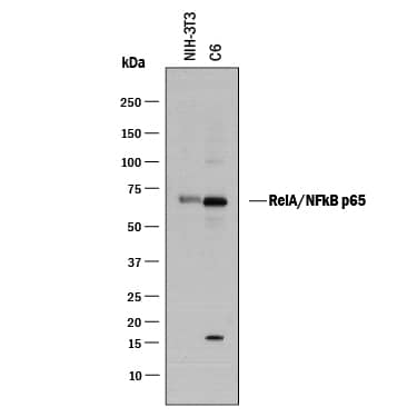
Detection of Mouse and Rat RelA/NF kappa B p65 by Western Blot. Western blot shows lysates of NIH-3T3 mouse embryonic fibroblast cell line and C6 rat glioma cell line. PVDF membrane was probed with 1 µg/mL of Sheep Anti-Human/Mouse RelA/NF kappa B p65 Antigen Affinity-purified Polyclonal Antibody (Catalog # AF5078) followed by HRP-conjugated Anti-Sheep IgG Secondary Antibody (Catalog # HAF016). A specific band was detected for RelA/NF kappa B p65 at approximately 65 kDa (as indicated). This experiment was conducted under reducing conditions and using Immunoblot Buffer Group 1.
 View Larger
Detection of RelA/NF kappa B p65-regulated Genes by Chromatin Immuno-precipitation. Jurkat human acute T cell leukemia cell line treated with 50 ng/mL PMA and 200 ng/mL calcium ionomycin for overnight was fixed using formaldehyde, resuspended in lysis buffer, and sonicated to shear chromatin. RelA/NF kappa B p65/DNA complexes were immunoprecipitated using 5 µg Sheep Anti-Human/Mouse/Rat RelA/NF kappa B p65 Antigen Affinity-purified Polyclonal Antibody (Catalog # AF5078) or control antibody (Catalog # 5-001-A) for 15 minutes in an ultrasonic bath, followed by Biotinylated Anti-Sheep IgG Secondary Antibody (Catalog # [[catalogNumber:BAF016)]]. Immunocomplexes were captured using 50 µL of MagCellect Streptavidin Ferrofluid (Catalog # [[catalogNumber:MAG999]]) and DNA was purified using chelating resin solution. Thep21promoter was detected by standard PCR.
 View Larger
RelA/NF kappa B p65 in HeLa Human Cell Line. RelA/NF kappa B p65 was detected in immersion fixed HeLa human cervical epithelial carcinoma cells untreated (upper panel) or treated (lower panel) with 20 ng/mL Recombinant Human TNF-alpha (Catalog # 210-TA) for 10 minutes using Sheep Anti-Human/Mouse/Rat RelA/NF kappa B p65 Antigen Affinity-purified Polyclonal Antibody (Catalog # AF5078) at 10 µg/mL for 3 hours at room temperature. Cells were stained using the NorthernLights™ 557-conjugated Anti-Sheep IgG Secondary Antibody (red; Catalog # NL010) and counterstained with DAPI (blue). Specific staining was localized to cytoplasm in untreated cells and nuclei in treated cells. View our protocol for Fluorescent ICC Staining of Cells on Coverslips.
 View Larger
RelA/NF kappa B p65 in Human Squamous Cell Carcinoma. RelA/NF kappa B p65 was detected in immersion fixed paraffin-embedded sections of human squamous cell carcinoma using Sheep Anti-Human/Mouse/Rat RelA/NF kappa B p65 Antigen Affinity-purified Polyclonal Antibody (Catalog # AF5078) at 3 µg/mL overnight at 4 °C. Tissue was stained using the Anti-Sheep HRP-DAB Cell & Tissue Staining Kit (brown; Catalog # CTS019) and counterstained with hematoxylin (blue). Specific staining was localized to cytoplasm in cancer cells. View our protocol for Chromogenic IHC Staining of Paraffin-embedded Tissue Sections.
 View Larger
Detection of Human, Mouse, and Rat RelA/NF kappa B p65 by Simple WesternTM. Simple Western lane view shows lysates of HeLa human cervical epithelial carcinoma cell line, Neuro-2A mouse neuroblastoma cell line, and C6 rat glioma cell line, loaded at 0.5 mg/mL. A specific band was detected for RelA/NF kappa B p65 at approximately 65 kDa (as indicated) using 20 µg/mL of Sheep Anti-Human/Mouse/Rat RelA/NF kappa B p65 Antigen Affinity-purified Polyclonal Antibody (Catalog # AF5078) followed by 1:50 dilution of HRP-conjugated Anti-Sheep IgG Secondary Antibody (Catalog # HAF016). This experiment was conducted under reducing conditions and using the 12-230 kDa separation system.
 View Larger
Western Blot Shows Human RelA/NF kappa B p65 Specificity by Using Knockout Cell Line. Western blot shows lysates of HeLa human cervical epithelial carcinoma parental cell line and RelA/NF kappa B p65 knockout HeLa cell line (KO). PVDF membrane was probed with 1 µg/mL of Sheep Anti-Human/Mouse/Rat RelA/NF kappa B p65 Antigen Affinity-purified Polyclonal Antibody (Catalog # AF5078) followed by HRP-conjugated Anti-Sheep IgG Secondary Antibody (Catalog # HAF016). A specific band was detected for RelA/NF kappa B p65 at approximately 65 kDa (as indicated) in the parental HeLa cell line, but is not detectable in knockout HeLa cell line. GAPDH (Catalog # AF5718) is shown as a loading control. This experiment was conducted under reducing conditions and using Immunoblot Buffer Group 1.
Reconstitution Calculator
Preparation and Storage
- 12 months from date of receipt, -20 to -70 °C as supplied.
- 1 month, 2 to 8 °C under sterile conditions after reconstitution.
- 6 months, -20 to -70 °C under sterile conditions after reconstitution.
Background: RelA/NFkB p65
RelA belongs to a family of transcription factors (NF kappa B (nuclear factor kappa from B cells) complex) that play a fundamental role in inflammatory and immune responses. The NF kappa B complex is composed of a heterodimer of a Rel family member (RelA, c-Rel, RelB) and either NF kappa B1 or NF kappa B2 subunits. RelA and NF kappa B1 are the most common heterodimeric pair. The NF kappa B complex is sequestered in the cytoplasm by inhibitory I kappa B proteins. Upon cellular activation, the ubiquitin-proteosome pathway degrades the I kappa B proteins allowing the NF kappa B complex to translocate to the nucleus and activate gene transcription.
Product Datasheets
Citations for Human/Mouse/Rat RelA/NF kappa B p65 Antibody
R&D Systems personnel manually curate a database that contains references using R&D Systems products. The data collected includes not only links to publications in PubMed, but also provides information about sample types, species, and experimental conditions.
3
Citations: Showing 1 - 3
Filter your results:
Filter by:
-
Anagliptin prevents lipopolysaccharide (LPS)- induced inflammation and activation of macrophages
Authors: F Yu, W Tian, J Dong
International immunopharmacology, 2022-01-16;104(0):108514.
Species: Mouse
Sample Types: Cell Lysates
Applications: Western Blot -
TET repression and increased DNMT activity synergistically induce aberrant DNA methylation
Authors: H Takeshima, T Niwa, S Yamashita, T Takamura-E, N Iida, M Wakabayash, S Nanjo, M Abe, T Sugiyama, YJ Kim, T Ushijima
J. Clin. Invest., 2020-10-01;0(0):.
Species: Human
Sample Types: Cell Lysates
Applications: Immunoprecipitation -
Characterization of short range DNA looping in endotoxin-mediated transcription of the murine inducible nitric-oxide synthase (iNOS) gene.
Authors: Guo H, Mi Z, Kuo PC
J. Biol. Chem., 2008-07-02;283(37):25209-17.
Species: Mouse
Sample Types: Cell Lysates
Applications: ChIP, Western Blot
FAQs
No product specific FAQs exist for this product, however you may
View all Antibody FAQsReviews for Human/Mouse/Rat RelA/NF kappa B p65 Antibody
Average Rating: 5 (Based on 2 Reviews)
Have you used Human/Mouse/Rat RelA/NF kappa B p65 Antibody?
Submit a review and receive an Amazon gift card.
$25/€18/£15/$25CAN/¥75 Yuan/¥2500 Yen for a review with an image
$10/€7/£6/$10 CAD/¥70 Yuan/¥1110 Yen for a review without an image
Filter by:


