Human/Mouse Semaphorin 6B Antibody Summary
Leu26-Ser603
Accession # Q9H3T3
Applications
Recombinant Mouse Semaphorin 6B
Please Note: Optimal dilutions should be determined by each laboratory for each application. General Protocols are available in the Technical Information section on our website.
Scientific Data
 View Larger
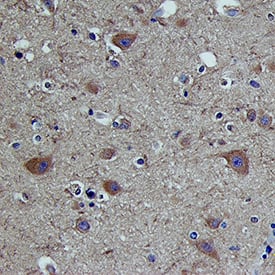
Detection of Semaphorin 6B in Human Brain Cortex. Semaphorin 6B was detected in immersion fixed paraffin-embedded sections of Human Brain Cortex using Goat Anti-Human/Mouse Semaphorin 6B Antigen Affinity-purified Polyclonal Antibody (Catalog # AF2094) at 15 µg/mL for 1 hour at room temperature followed by incubation with the Anti-Goat IgG VisUCyte™ HRP Polymer Antibody (Catalog # VC004). Before incubation with the primary antibody, tissue was subjected to heat-induced epitope retrieval using VisUCyte Antigen Retrieval Reagent-Basic (Catalog # VCTS021). Tissue was stained using DAB (brown) and counterstained with hematoxylin (blue). Specific staining was localized to membrane and cytoplasm in neurons. View our protocol for IHC Staining with VisUCyte HRP Polymer Detection Reagents.
Reconstitution Calculator
Preparation and Storage
- 12 months from date of receipt, -20 to -70 °C as supplied.
- 1 month, 2 to 8 °C under sterile conditions after reconstitution.
- 6 months, -20 to -70 °C under sterile conditions after reconstitution.
Background: Semaphorin 6B
Semaphorin 6B (Sema6B) is a 120 kDa member of the Semaphorin family of axon guidance molecules (1‑4). The four known Class 6 semaphorins are type I transmembrane glycoproteins with Sema domains but without other domains, making them most like Class 1 invertebrate semaphorins in structure (1‑4). Amino acid (aa) identity of Class 6 semaphorins is around 40% overall, but 53‑64% within the Sema domain. Sema6B is expressed developmentally in subregions of the nervous system and muscle, and at low levels in most adult tissues (3, 4). Human Sema6B cDNA encodes a 25 aa signal sequence, a 579 aa extracellular domain (ECD) including the Sema domain, a 20 aa transmembrane sequence and a 263 aa cytoplasmic portion. A cytoplasmic proline-rich sequence interacts with the SH3 domain of the c-src signaling protein (4). Full-length Sema6B is thought to form disulfide-linked homodimers (4). Alternate exon splicing creates a 492 aa (presumably) secreted form (Sema6B.1), and a 657 aa form with a shortened cytoplasmic tail (Sema6B.2) (3). Human Sema6B ECD shows 94%, 94%, 96% and 89% aa identity with corresponding mouse, rat, bovine and canine sequences, respectively. Crystal structures of semaphorins reveal that the 500 aa Sema domain forms an integrin-like seven-blade beta -propeller structure stabilized by 14 conserved cysteine residues (5). Sema6B is highly expressed in some glioblastoma and breast cancer cell lines. All-trans retinoic acid slows cancer cell growth and down‑regulates Sema6B expression, probably via dimerization with peroxisome proliferator-activated receptors (PPAR) that have a response element on the Sema6B gene (3, 6, 7). Semaphorins transduce signals through transmembrane plexins, either directly, or by binding associated neuropilin receptors. Plexin-A4 binds Sema6A (high affinity) and 6B (low affinity) and mediates sympathetic ganglion axon-repulsion, independent of neuropilin-1 (8).
- Neufeld, G. et al. (2005) Front. Biosci. 10:751.
- Chedotal, A. et al. (2005) Cell Death Differ. 12:1044.
- Correa, R. G. et al. (2001) Genomics 73:343.
- Eckhardt, F. et al. (1997) Mol. Cell. Neurosci. 9:409.
- Gherardi, E. et al. (2004) Curr. Opin. Struct. Biol. 14:669.
- Collet, P. et al. (2004) Genomics 83:1141.
- Murad, H. et al. (2006) Int. J. Oncol. 28:977.
- Suto, F. et al. (2005) J. Neurosci. 25:3628.
Product Datasheets
Citations for Human/Mouse Semaphorin 6B Antibody
R&D Systems personnel manually curate a database that contains references using R&D Systems products. The data collected includes not only links to publications in PubMed, but also provides information about sample types, species, and experimental conditions.
2
Citations: Showing 1 - 2
Filter your results:
Filter by:
-
Recognition of Semaphorin Proteins by P. sordellii Lethal Toxin Reveals Principles of Receptor Specificity in Clostridial Toxins
Authors: Hunsang Lee, Greg L. Beilhartz, Iga Kucharska, Swetha Raman, Hong Cui, Mandy Hiu Yi Lam et al.
Cell
-
Roles of semaphorin-6B and plexin-A2 in lamina-restricted projection of hippocampal mossy fibers.
Authors: Tawarayama H, Yoshida Y, Suto F, Mitchell KJ, Fujisawa H
J. Neurosci., 2010-05-19;30(20):7049-60.
Species: Mouse
Sample Types: Tissue Homogenates
Applications: Western Blot
FAQs
No product specific FAQs exist for this product, however you may
View all Antibody FAQsReviews for Human/Mouse Semaphorin 6B Antibody
There are currently no reviews for this product. Be the first to review Human/Mouse Semaphorin 6B Antibody and earn rewards!
Have you used Human/Mouse Semaphorin 6B Antibody?
Submit a review and receive an Amazon gift card.
$25/€18/£15/$25CAN/¥75 Yuan/¥2500 Yen for a review with an image
$10/€7/£6/$10 CAD/¥70 Yuan/¥1110 Yen for a review without an image

