Human/Mouse Serpin F1/PEDF Antibody Summary
Asp44-Pro418
Accession # P36955
Applications
Please Note: Optimal dilutions should be determined by each laboratory for each application. General Protocols are available in the Technical Information section on our website.
Scientific Data
 View Larger
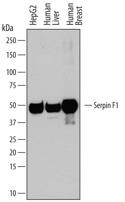
Detection of Human Serpin F1/PEDF by Western Blot. Western blot shows lysates of HepG2 human hepatocellular carcinoma cell line, human liver tissue, and human breast tissue. PVDF membrane was probed with 0.2 µg/mL of Goat Anti-Human/Mouse Serpin F1/PEDF Antigen Affinity-purified Polyclonal Antibody (Catalog # AF1177) followed by HRP-conjugated Anti-Goat IgG Secondary Antibody (Catalog # HAF109). A specific band was detected for Serpin F1/PEDF at approximately 50 kDa (as indicated). This experiment was conducted under reducing conditions and using Immunoblot Buffer Group 1.
 View Larger
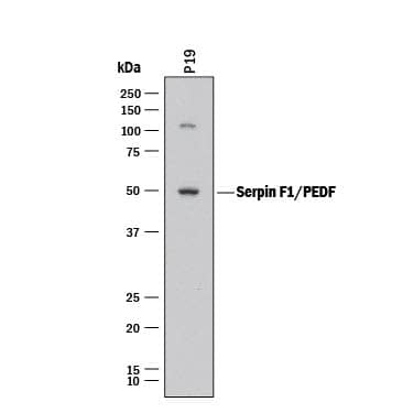
Detection of Mouse Serpin F1/PEDF by Western Blot. Western blot shows lysates of P19 mouse embryonal carcinoma cell line. PVDF membrane was probed with 2 µg/mL of Goat Anti-Human/Mouse Serpin F1/PEDF Antigen Affinity-purified Polyclonal Antibody (Catalog # AF1177) followed by HRP-conjugated Anti-Goat IgG Secondary Antibody (Catalog # HAF017). A specific band was detected for Serpin F1/PEDF at approximately 50 kDa (as indicated). This experiment was conducted under reducing conditions and using Immunoblot Buffer Group 1.
 View Larger
Serpin F1/PEDF in Human Kidney. Serpin F1/PEDF was detected in immersion fixed paraffin-embedded sections of human kidney using Goat Anti-Human/Mouse Serpin F1/PEDF Antigen Affinity-purified Polyclonal Antibody (Catalog # AF1177) at 15 µg/mL overnight at 4 °C. Tissue was stained using the Anti-Goat HRP-DAB Cell & Tissue Staining Kit (brown; Catalog # CTS008) and counterstained with hematoxylin (blue). Specific staining was localized to convoluted tubules. View our protocol for Chromogenic IHC Staining of Paraffin-embedded Tissue Sections.
 View Larger
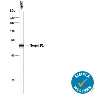
Detection of Human Serpin F1/PEDF by Simple WesternTM. Simple Western lane view shows lysates of HepG2 human hepatocellular carcinoma cell line, loaded at 0.2 mg/mL. A specific band was detected for Serpin F1/PEDF at approximately 56 kDa (as indicated) using 2 µg/mL of Goat Anti-Human/Mouse Serpin F1/PEDF Antigen Affinity-purified Polyclonal Antibody (Catalog # AF1177) followed by 1:50 dilution of HRP-conjugated Anti-Goat IgG Secondary Antibody (Catalog # HAF109). This experiment was conducted under reducing conditions and using the 12-230 kDa separation system.
Reconstitution Calculator
Preparation and Storage
- 12 months from date of receipt, -20 to -70 °C as supplied.
- 1 month, 2 to 8 °C under sterile conditions after reconstitution.
- 6 months, -20 to -70 °C under sterile conditions after reconstitution.
Background: Serpin F1/PEDF
Serpin F1 (serine proteinase inhibitor-clade F #1; also PEDF, PIG35 and EPC-1) is a secreted, 50 kDa glycoprotein member of the clade F-subfamily, serpin superfamily of protease inhibitors. It is expressed by diverse cell types such as retinal pigment and breast epithelium, fibroblasts, astrocytes and hepatocytes. Serpin F1 circulates in blood and binds to type I collagen plus heparan sulfate. Although it is a serpin, it belongs to a class of serpins that are nonprotease inhibiting, and instead is known to possess potent antiangiogenic and neurotrophic activity. Human Serpin F1 is 418 amino acids (aa) in length. It contains a 19 aa signal sequence plus a 399 aa mature region that shows an NLS (aa 146-149), a neuroprotective motif (aa 354-359) and an antiangiogenesis segment (aa 387‑411). Phosphorylation occurs at multiple sites that affect bioactivity. Over aa 44‑418, human Serpin F1 shares 87% aa identity with mouse Serpin F1.
Product Datasheets
Citations for Human/Mouse Serpin F1/PEDF Antibody
R&D Systems personnel manually curate a database that contains references using R&D Systems products. The data collected includes not only links to publications in PubMed, but also provides information about sample types, species, and experimental conditions.
9
Citations: Showing 1 - 9
Filter your results:
Filter by:
-
Pigment Epithelium Derived Factor Is Involved in the Late Phase of Osteosarcoma Metastasis by Increasing Extravasation and Cell-Cell Adhesion
Authors: Sei Kuriyama, Gentaro Tanaka, Kurara Takagane, Go Itoh, Masamitsu Tanaka
Frontiers in Oncology
-
Pigment epithelium-derived factor mediates retinal ganglion cell neuroprotection by suppression of caspase-2
Authors: V Vigneswara, Z Ahmed
Cell Death Dis, 2019-02-04;10(2):102.
Species: Rat
Sample Types: Whole Cells
Applications: Agonist Activity -
HtrA1 Mediated Intracellular Effects on Tubulin Using a Polarized RPE Disease Model
Authors: E Melo, P Oertle, C Trepp, H Meisterman, T Burgoyne, L Sborgi, AC Cabrera, CY Chen, JC Hoflack, T Kam-Thong, R Schmucki, L Badi, N Flint, ZE Ghiani, F Delobel, C Stucki, G Gromo, A Einhaus, B Hornsperge, S Golling, J Siebourg-P, F Gerber, B Bohrmann, C Futter, T Dunkley, S Hiller, O Schilling, V Enzmann, S Fauser, M Plodinec, R Iacone
EBioMedicine, 2017-12-13;0(0):.
Species: Human
Sample Types: Cell Lysates, Whole Cells
Applications: ICC, Western Blot -
Quantitative changes in the protein and miRNA cargo of plasma exosome-like vesicles after exposure to ionizing radiation
Authors: R Yentrapall, J Merl-Pham, O Azimzadeh, L Mutschelkn, C Peters, SM Hauck, MJ Atkinson, S Tapio, S Moertl
Int. J. Radiat. Biol., 2017-03-07;0(0):1-12.
Species: Human
Sample Types: Cell Lysates
Applications: Western Blot -
Pigment epithelium-derived factor (PEDF) binds to caveolin-1 and inhibits the pro-inflammatory effects of caveolin-1 in endothelial cells.
Authors: Matsui T, Higashimoto Y, Taira J, Yamagishi S
Biochem Biophys Res Commun, 2013-10-23;441(2):405-10.
Species: Human
Sample Types: Cell Lysates
Applications: Western Blot -
Secretome and degradome profiling shows that Kallikrein-related peptidases 4, 5, 6, and 7 induce TGFbeta-1 signaling in ovarian cancer cells.
Authors: Shahinian H, Loessner D, Biniossek M, Kizhakkedathu J, Clements J, Magdolen V, Schilling O
Mol Oncol, 2013-10-01;8(1):68-82.
Species: Human
Sample Types: Cell Culture Supernates
Applications: Western Blot -
Long-Term Retinal PEDF Overexpression Prevents Neovascularization in a Murine Adult Model of Retinopathy.
Authors: Haurigot V, Villacampa P, Ribera A, Bosch A, Ramos D, Ruberte J, Bosch F
PLoS ONE, 2012-07-20;7(7):e41511.
Species: Mouse
Sample Types: Tissue Homogenates, Whole Tissue
Applications: IHC-P, Western Blot -
A therapeutic strategy for choroidal neovascularization based on recruitment of mesenchymal stem cells to the sites of lesions.
Authors: Hou HY, Liang HL, Wang YS
Mol. Ther., 2010-07-20;18(10):1837-45.
Species: Mouse
Sample Types: Whole Tissue
Applications: IHC-Fr -
Inhibition of nuclear translocation of apoptosis-inducing factor is an essential mechanism of the neuroprotective activity of pigment epithelium-derived factor in a rat model of retinal degeneration.
Authors: Murakami Y, Ikeda Y, Yonemitsu Y, Onimaru M, Nakagawa K, Kohno R, Miyazaki M, Hisatomi T, Nakamura M, Yabe T, Hasegawa M, Ishibashi T, Sueishi K
Am. J. Pathol., 2008-10-09;173(5):1326-38.
Species: Human
Sample Types: Recombinant Protein
Applications: Neutralization
FAQs
No product specific FAQs exist for this product, however you may
View all Antibody FAQsReviews for Human/Mouse Serpin F1/PEDF Antibody
There are currently no reviews for this product. Be the first to review Human/Mouse Serpin F1/PEDF Antibody and earn rewards!
Have you used Human/Mouse Serpin F1/PEDF Antibody?
Submit a review and receive an Amazon gift card.
$25/€18/£15/$25CAN/¥75 Yuan/¥2500 Yen for a review with an image
$10/€7/£6/$10 CAD/¥70 Yuan/¥1110 Yen for a review without an image
