Human/Mouse SorCS3 Antibody Summary
Ala131-Ser1122
Accession # Q8VI51
Applications
Please Note: Optimal dilutions should be determined by each laboratory for each application. General Protocols are available in the Technical Information section on our website.
Scientific Data
 View Larger
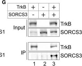
Detection of Chinese hamster SorCS3 by Western Blot Altered subcellular localization of TrkB in primary neurons and brains from S1/3 KO miceAWorkflow of the cell surface proteome analysis in primary neurons.BResults of quantitative label‐free proteomics comparing the surface proteomes of WT and S1/3 KO primary cortical neurons. Plot represents –log10(P‐value) and log2 (relative levels S1/3KO/WT) obtained for each protein. For proteins that were detected in either S1/3 KO or WT samples only, the log2(S1/3 KO/WT) was set to 10 or ‐10, respectively. Threshold values are 1.3 for –log10(P‐value) and ±0.3 for log2(S1/3 KO/WT). Proteins showing higher (green) or lower (red) surface levels in S1/3 KO as compared to WT neurons are indicated. Proteins with unchanged cell surface levels are indicated in blue. n = 3 biological replicates/group (with each biological replicate run in two technical replicates).CScheme of brain membrane fractionation used to analyze subcellular localization of TrkB in vivo (see method section for details).DRepresentative Western blotting of TrkB in various brain membrane fractions obtained as described in panel (C). Highlighted lanes represent P3 and P2 fractions, quantified in (E). Detection of PSD95, synaptophysin, and Golgin 97 was used to assess the accuracy of subcellular fractionation.EQuantification of total TrkB distribution between P3 (light membrane fraction) and P2 (synaptosomal fraction) from Western blots exemplified in panel (D) (n = 5 mice/group, 7–12 weeks of age). The TrkB pool is shifted toward the light membrane (P3) fraction in S1/3 KO brains. Data are shown as mean ± SEM and analyzed using two‐way ANOVA with Bonferroni post‐test. ****P < 0.0001.F, GCo‐immunoprecipitation of SORCS1 and SORCS3 with TrkB. TrkB was immunoprecipitated from transiently transfected Chinese hamster ovary cells and the presence of SORCS1 or SORCS3 in the immunoprecipitates documented by Western blotting. Panel input shows levels of the indicated proteins in the cell lysates prior to immunoprecipitation. Panel IP documents co‐immunoprecipitation of SORCS1 (F) and SORCS3 (G) from cells expressing (lanes 3) but not from cells lacking TrkB (lanes 2). The experiment was replicated twice.Source data are available online for this figure. Image collected and cropped by CiteAb from the following publication (https://pubmed.ncbi.nlm.nih.gov/29440124), licensed under a CC-BY license. Not internally tested by R&D Systems.
Reconstitution Calculator
Preparation and Storage
- 12 months from date of receipt, -20 to -70 °C as supplied.
- 1 month, 2 to 8 °C under sterile conditions after reconstitution.
- 6 months, -20 to -70 °C under sterile conditions after reconstitution.
Background: SorCS3
SorCS3 is a type I transmembrane receptor of the mammalian Vps10p (vacuolar protein‑sorting 10 protein) family of receptors that includes sortilin, SorLA, and three SorCS proteins (1, 2). It is synthesized as an ~1220 amino acid (aa) preproform with a 33 aa signal sequence and a 100 aa propeptide. After proteolytic release of the propeptide at a furin‑type consensus sequence, the mature SorCS3 is a 100‑110 kDa transmembrane protein with a 992 aa extracellular/lumenal domain (ECD). Mouse and human SorCS3 ECD share 92% aa sequence identity. They also share ~70% and ~45% aa identity with SorCS1 and SorCS2 ECDs, respectively. The ECD contains an imperfect leucine‑rich repeat (LRR) and a Vps10p domain that binds both pro- and mature NGF (2, 3). The metalloproteinase TACE/ADAM17 is able to cleave SorCS3 near the membrane either constitutively, or at an increased rate when induced by phorbol esters (4). The shed ECD is able to bind PDGF‑BB and the NGF propeptide (4). Unlike sortilin, the SorCS3 propeptide has no known function; it does not block NGF binding or propeptide cleavage (3, 5). SorCS3 is predominantly expressed on the plasma membrane (3). It can be slowly internalized but, despite the presence of a sorting domain, there is no evidence for SorCS3‑mediated intracellular trafficking activity (3). It is expressed in the embryonic and adult central nervous system in areas distinct from that of SorCS1 and SorCS2 (1). Neuronal activity upregulates SorCS3 expression in the hippocampus (1).
- Hermey, G. et al. (2004) J. Neurochem. 88:1470.
- Hampe, W. et al. (2001) Hum. Genet. 108:529.
- Westergaard, U. B. et al. (2005) FEBS Lett. 579:1172.
- Hermey, G. et al. (2006) Biochem. J. 395:285.
- Westergaard, U. et al. (2004) J. Biol. Chem. 279:50221.
Product Datasheets
Citations for Human/Mouse SorCS3 Antibody
R&D Systems personnel manually curate a database that contains references using R&D Systems products. The data collected includes not only links to publications in PubMed, but also provides information about sample types, species, and experimental conditions.
4
Citations: Showing 1 - 4
Filter your results:
Filter by:
-
Neuronal cadherin (NCAD) increases sensory neurite formation and outgrowth on astrocytes
Authors: Toby A. Ferguson, Steven S. Scherer
Neuroscience Letters
-
SORCS1 and SORCS3 control energy balance and orexigenic peptide production
Authors: A Subkhangul, AR Malik, G Hermey, O Popp, G Dittmar, T Rathjen, MN Poy, A Stumpf, PS Beed, D Schmitz, T Breiderhof, TE Willnow
EMBO Rep., 2018-02-12;19(4):.
Species: Mouse
Sample Types: Cell Lysates
Applications: Western Blot -
Prions amplify through degradation of the VPS10P sorting receptor sortilin
Authors: K Uchiyama, M Tomita, M Yano, J Chida, H Hara, NR Das, A Nykjaer, S Sakaguchi
PLoS Pathog., 2017-06-30;13(6):e1006470.
Species: Mouse
Sample Types: Cell Lysates
Applications: Western Blot -
A synthetic connexin 43 mimetic peptide augments corneal wound healing.
Authors: Moore K, Bryant ZJ, Ghatnekar G et al.
Exp Eye Res.
FAQs
No product specific FAQs exist for this product, however you may
View all Antibody FAQsReviews for Human/Mouse SorCS3 Antibody
There are currently no reviews for this product. Be the first to review Human/Mouse SorCS3 Antibody and earn rewards!
Have you used Human/Mouse SorCS3 Antibody?
Submit a review and receive an Amazon gift card.
$25/€18/£15/$25CAN/¥75 Yuan/¥2500 Yen for a review with an image
$10/€7/£6/$10 CAD/¥70 Yuan/¥1110 Yen for a review without an image

