Human PD-L2/B7-DC Antibody Summary
Leu20-Pro219
Accession # Q9BQ51
Applications
Please Note: Optimal dilutions should be determined by each laboratory for each application. General Protocols are available in the Technical Information section on our website.
Scientific Data
 View Larger
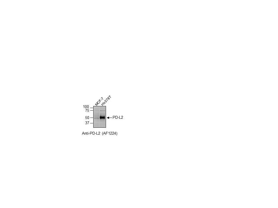
Detection of Human PD‑L2/B7‑DC by Western Blot. Western blot shows lysates of human lung tissue and HDLM-2 human Hodgkin's lymphoma cell line. PVDF membrane was probed with 2 µg/mL of Goat Anti-Human PD-L2/B7-DC Antigen Affinity-purified Polyclonal Antibody (Catalog # AF1224) followed by HRP-conjugated Anti-Goat IgG Secondary Antibody (Catalog # HAF017). Specific bands were detected for PD-L2/B7-DC at approximately 45-50 kDa (as indicated). This experiment was conducted under reducing conditions and using Immunoblot Buffer Group 1.
 View Larger
Detection of Human PD-L2/B7-DC/PDCD1LG2 by Immunohistochemistry PD-L1, PD-L2, PD-1, CD8, and CD4 expression in p16-positive and p16-negative HNSCC. PD-L1, PD-L2, PD-1, CD8, and CD4 expression was assessed in tumor biopsy tissue from five p16-positive and four p16-negative HNSCC patients using immuno-histochemistry (details in Methods). Representative staining (scale bars, 100 μm) and cumulative data of marker expression (grading scale PD-L1/PD-L2: 1, low; 2, moderate; 3 high expression; grading scale PD-1, CD8, CD4: 1, <50 cells/field; 2, 50–150 cells/field; 3, >150 cells/field). Image collected and cropped by CiteAb from the following open publication (https://pubmed.ncbi.nlm.nih.gov/31379843), licensed under a CC-BY license. Not internally tested by R&D Systems.
Reconstitution Calculator
Preparation and Storage
- 12 months from date of receipt, -20 to -70 °C as supplied.
- 1 month, 2 to 8 °C under sterile conditions after reconstitution.
- 6 months, -20 to -70 °C under sterile conditions after reconstitution.
Background: PD-L2/B7-DC
T cells require a signal induced by the engagement of the T cell receptor and a “co‑stimulatory” signal(s) through distinct T cell surface molecules for optimal T cell activation and tolerance. Members of the B7 superfamily of counter-receptors were identified by their ability to interact with co‑stimulatory molecules found on the surface of T cells. Members of the B7 superfamily include B7-1 (CD80), B7-2 (CD86), B7-H1 (PD-L1), B7-H2 (B7RP-1), B7-H3, and PD-L2 (B7-DC) (1). B7 proteins are immunoglobulin (Ig) superfamily members with extracellular Ig-V-like and Ig-C-like domains and short cytoplasmic domains. Among the family members, they share from 20‑40% amino acid (aa) sequence identity. The cloned human PD-L2 cDNA encodes a 273 aa type I membrane precursor protein with a putative 20 aa signal peptide, a 201 aa extracellular region containing one V-like and one C-like Ig domain, a 24 aa transmembrane region, and a 28 aa cytoplasmic domain. The extracellular domains of mouse and human PD-L2 share approximately 70% aa sequence identity (2). PD-L2 is one of two ligands for programmed death-1 (PD-1), a member of the CD28 family of immuno-receptors. The other identified ligand is PD-L1. Human PD-L1 and PD-L2 share approximately 41% aa sequence identity and have similar functions. PD-L2 is broadly expressed in tissues. Highest expression was detected by Northern blot analysis in heart, placenta, liver, pancreas, spleen, and lymph node. Lower amounts of expression were observed in lung, smooth muscle, and thymus. Expression of PD-L2 on antigen presenting cell has been examined in detail. Resting B cells, monocytes and dendritic cells do not express PD-L2, expression however can be induced by LPS or BCR activation in B cells, INF-gamma treatment in monocytes, or LPS plus IFN-gamma treatment of dendritic cells. PD-L2 expression is also up regulated in a variety of tumor cell lines. On previously activated T cells, PD-L2 interaction with PD-1 inhibits TCR-mediated proliferation and cytokine production, suggesting an inhibitory role in regulating immune responses. In contrast, a co‑stimulatory function for the PD-L2 on resting T cells activated with sub-optimal TCR signals has also been reported (3).
- Coyle, A.J. and J-C. Gutierrrez-Ramos (2001) Nature Immunol. 2:203.
- Latchman Y. et al. (2001) Nature Immun. 2:261.
- Carreno, B.M. and M. Collins (2002) Annu. Rev. Immunol. 20:29.
Product Datasheets
Citations for Human PD-L2/B7-DC Antibody
R&D Systems personnel manually curate a database that contains references using R&D Systems products. The data collected includes not only links to publications in PubMed, but also provides information about sample types, species, and experimental conditions.
7
Citations: Showing 1 - 7
Filter your results:
Filter by:
-
TLR9 Mediated Tumor-Stroma Interactions in Human Papilloma Virus (HPV)-Positive Head and Neck Squamous Cell Carcinoma Up-Regulate PD-L1 and PD-L2
Authors: Paramita Baruah, Jessica Bullenkamp, Philip O. G. Wilson, Michael Lee, Juan Carlos Kaski, Ingrid E. Dumitriu
Frontiers in Immunology
-
Immune response in breast cancer brain metastases and their microenvironment: the role of the PD-1/PD-L axis.
Authors: Duchnowska R, Peksa R, Radecka B et al.
Breast Cancer Res
-
Currently Used Laboratory Methodologies for Assays Detecting PD-1, PD-L1, PD-L2 and Soluble PD-L1 in Patients with Metastatic Breast Cancer
Authors: S Jeong, N Lee, MJ Park, K Jeon, W Song
Cancers, 2021-10-18;13(20):.
Species: Human
Sample Types: Whole Tissue
Applications: IHC -
Human amnion-derived mesenchymal stem cells attenuate xenogeneic graft-versus-host disease by preventing T cell activation and proliferation
Authors: Y Tago, C Kobayashi, M Ogura, J Wada, S Yamaguchi, T Yamaguchi, M Hayashi, T Nakaishi, H Kubo, Y Ueda
Scientific Reports, 2021-01-28;11(1):2406.
Species: Human
Sample Types: Whole Cells
Applications: Neutralization -
Mesenchymal Stromal Cell Secretion of Programmed Death-1 Ligands Regulates T Cell Mediated Immunosuppression
Stem Cells, 2016-10-26;0(0):.
Species: Human
Sample Types: Whole Cells
Applications: Immunoprecipitation -
PD-L1, PD-L2 and PD-1 expression in metastatic melanoma: Correlation with tumor-infiltrating immune cells and clinical outcome
Authors: Joseph M Obeid
Oncoimmunology, 2016-09-20;5(11):e1235107.
Species: Human
Sample Types: Whole Tissue
Applications: IHC-P -
12-O-tetradecanoyl phorbol 13-acetate induces the expression of B7-DC, -H1, -H2, and -H3 in K562 cells.
Authors: Jang BC, Park YK, Choi IH, Kim SP, Hwang JB, Baek WK, Suh MH, Mun KC, Suh SI
Int. J. Oncol., 2007-12-01;31(6):1439-47.
Species: Human
Sample Types: Whole Cells
Applications: Flow Cytometry
FAQs
No product specific FAQs exist for this product, however you may
View all Antibody FAQsReviews for Human PD-L2/B7-DC Antibody
Average Rating: 5 (Based on 1 Review)
Have you used Human PD-L2/B7-DC Antibody?
Submit a review and receive an Amazon gift card.
$25/€18/£15/$25CAN/¥75 Yuan/¥2500 Yen for a review with an image
$10/€7/£6/$10 CAD/¥70 Yuan/¥1110 Yen for a review without an image
Filter by:


