Human Phospho-p70 S6 Kinase (T389) Antibody
Human Phospho-p70 S6 Kinase (T389) Antibody Summary
Applications
Please Note: Optimal dilutions should be determined by each laboratory for each application. General Protocols are available in the Technical Information section on our website.
Scientific Data
 View Larger
Detection of Human Phospho-p70 S6 Kinase (T389)/p85 S6 Kinase (T412) by Western Blot. Western blot shows lysates of MCF-7 human breast cancer cell line untreated (-) or treated (+) with 100 ng/mL Recombinant Human IGF-I (Catalog # 291-G1) for 60 minutes. PVDF membrane was probed with 0.1 µg/mL of Rabbit Anti-Human Phospho-p70 S6 Kinase (T389) Monoclonal Antibody (Catalog # MAB8963) followed by HRP-conjugated Anti-Rabbit IgG Secondary Antibody (Catalog # HAF008). Specific bands were detected for Phospho-p70 S6 Kinase (T389) and Phospho-p85 S6 Kinase (T412) at approximately 70 and 85 kDa, respectively (as indicated). This experiment was conducted under reducing conditions and using Immunoblot Buffer Group 1.
 View Larger
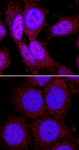
Phospho-p70 S6 Kinase (T389) in MCF‑7 Human Cell Line. p70 S6 Kinase phosphorylated at T389 was detected in immersion fixed serum starved MCF-7 human breast cancer cell line untreated (lower panel) or treated with Recombinant Human IGF-I (Catalog # 291-G1) using Rabbit Anti-Human Phospho-p70 S6 Kinase (T389) Monoclonal Antibody (Catalog # MAB8963) at 2 µg/mL for 3 hours at room temperature. Cells were stained using the NorthernLights™ 557-conjugated Anti-Rabbit IgG Secondary Antibody (red; Catalog # NL004) and counterstained with DAPI (blue). Specific staining was localized to cytoplasm and nuclei. View our protocol for Fluorescent ICC Staining of Cells on Coverslips.
 View Larger
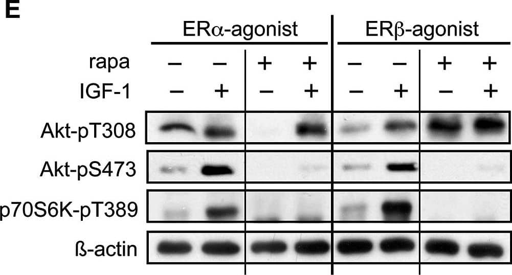
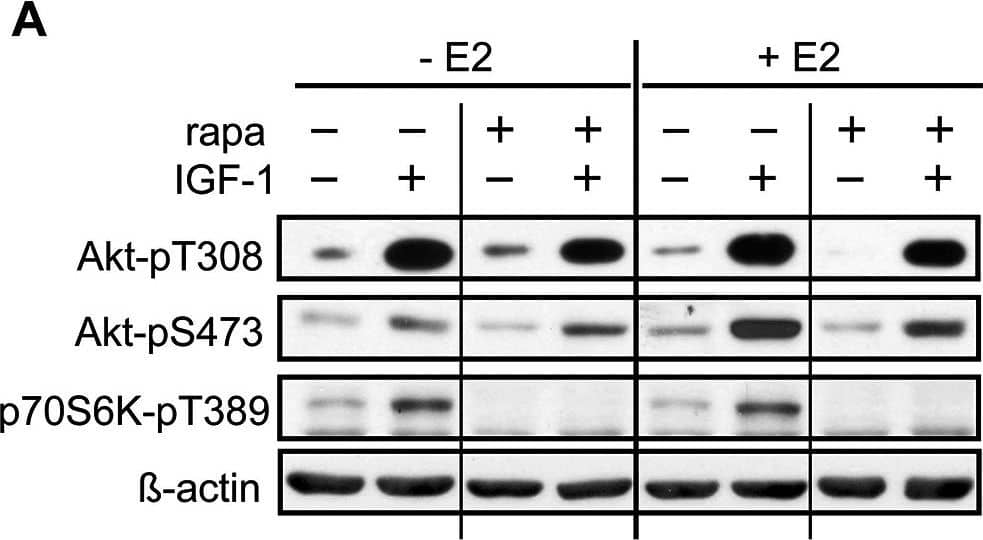
Detection of Mouse p70 S6 Kinase/S6K by Western Blot E2 regulates rapamycin effects on mTORC2 activity.A-H, Rapamycin lowers mTORC1 activity independent of presence of E2, ER alpha - or ER beta -agonist, however lowers mTORC2 activity dependent on presence of E2. A-D, HL-1 cells were grown to near confluence in medium containing 10 nM E2, and E-H, 10 nM ER alpha -agonist PPT or 1 nM ER beta -agonist DPN and serum starved for 24 hours prior to incubation with 20 nM rapamycin and IGF-1 for 24 h. A and E show representative westernblots. Equal loading was verified by blotting with antibodies against beta -actin or alpha -tubulin. For B,C,D and F,G,H, labeled bands were quantified with ImageJ software, normalized to loading and IGF-1 induced phosphorylation of indicated proteins was determined by ratio to the value of non-IGF-1 stimulated control cells. Mean ± SEM of fold stimulation by IGF-1 is shown of at least 3 independently performed experiments. * p < 0.05,** p < 0.009, *** p < 0.0001. If not indicated differently, significances are related to respective non-IGF-1 stimulated cells. Image collected and cropped by CiteAb from the following publication (https://dx.plos.org/10.1371/journal.pone.0123385), licensed under a CC-BY license. Not internally tested by R&D Systems.
Reconstitution Calculator
Preparation and Storage
- 12 months from date of receipt, -20 to -70 °C as supplied.
- 1 month, 2 to 8 °C under sterile conditions after reconstitution.
- 6 months, -20 to -70 °C under sterile conditions after reconstitution.
Background: p70 S6 Kinase
p70 S6 Kinase (p70S6K) is responsible for the phosphorylation of 40S ribosomal protein S6 and is ubiquitously expressed in human adult tissues (1). p70S6K is activated by serum stimulation and this activation is inhibited by wortmannin and rapamycin. p70S6K activity undergoes changes in the cell cycle and increases
20-fold in G1 cells released from G0 (2). p70S6K activation requires sequential phosphorylations at proline-directed residues in the putative autoinhibitory pseudosubstrate domain, as well as T389, a site phosphorylated by Phosphoinositide-Dependent Kinase 1 (PDK1).
- Ferrari, S. et al. (1994) Crit. Rev. Biochem. Mol. Biol. 29:385.
- Edelmann, H.M. et al. (1996) J. Biol. Chem. 271:963.
Product Datasheets
Citations for Human Phospho-p70 S6 Kinase (T389) Antibody
R&D Systems personnel manually curate a database that contains references using R&D Systems products. The data collected includes not only links to publications in PubMed, but also provides information about sample types, species, and experimental conditions.
6
Citations: Showing 1 - 6
Filter your results:
Filter by:
-
17-Beta-Estradiol Regulates mTORC2 Sensitivity to Rapamycin in Adaptive Cardiac Remodeling
Authors: Kusch A, Schmidt M, Gurgen D et al.
PLoS ONE
-
Radiotherapy orchestrates natural killer cell dependent antitumor immune responses through CXCL8
Authors: T Walle, JA Kraske, B Liao, B Lenoir, C Timke, E von Bohlen, F Tran, P Griebel, D Albrecht, A Ahmed, M Suarez-Car, A Jiménez-Sá, T Beikert, A Tietz-Dahl, AN Menevse, G Schmidt, M Brom, JHW Pahl, W Antonopoul, M Miller, RL Perez, F Bestvater, NA Giese, P Beckhove, P Rosenstiel, D Jäger, O Strobel, D Pe'er, N Halama, J Debus, A Cerwenka, PE Huber
Science Advances, 2022-03-23;8(12):eabh4050.
-
p85S6K sustains synaptic GluA1 to ameliorate cognitive deficits in Alzheimer’s disease
Authors: Jia-Bing Li, Xiao-Yu Hu, Mu-Wen Chen, Cai-Hong Xiong, Na Zhao, Yan-Hui Ge et al.
Translational Neurodegeneration
-
Characterizing the distributions of IDO-1 expressing macrophages/microglia in human and murine brains and evaluating the immunological and physiological roles of IDO-1 in RAW264.7/BV-2 cells
Authors: R Ji, L Ma, X Chen, R Sun, L Zhang, H Saiyin, W Wei
PLoS ONE, 2021-11-04;16(11):e0258204.
Species: Human
Sample Types: Cell Lysates
Applications: Western Blot -
Effect of gut microbiota on depressive-like behaviors in mice is mediated by the endocannabinoid system
Authors: G Chevalier, E Siopi, L Guenin-Mac, M Pascal, T Laval, A Rifflet, IG Boneca, C Demangel, B Colsch, A Pruvost, E Chu-Van, A Messager, F Leulier, G Lepousez, G Eberl, PM Lledo
Nature Communications, 2020-12-11;11(1):6363.
Species: Mouse
Sample Types: Cell Lysates
Applications: Western Blot -
WYE-354 restores Adriamycin sensitivity in multidrug-resistant acute myeloid leukemia cell lines
Authors: Sara M. Ibrahim, Sherin Bakhashab, Asad M. Ilyas, Peter N. Pushparaj, Sajjad Karim, Jalaluddin A. Khan et al.
Oncology Reports
FAQs
No product specific FAQs exist for this product, however you may
View all Antibody FAQsReviews for Human Phospho-p70 S6 Kinase (T389) Antibody
There are currently no reviews for this product. Be the first to review Human Phospho-p70 S6 Kinase (T389) Antibody and earn rewards!
Have you used Human Phospho-p70 S6 Kinase (T389) Antibody?
Submit a review and receive an Amazon gift card.
$25/€18/£15/$25CAN/¥75 Yuan/¥2500 Yen for a review with an image
$10/€7/£6/$10 CAD/¥70 Yuan/¥1110 Yen for a review without an image


