Human Phospho-STAT2 (Y690) Antibody Summary
*Small pack size (-SP) is supplied either lyophilized or as a 0.2 µm filtered solution in PBS.
Applications
Please Note: Optimal dilutions should be determined by each laboratory for each application. General Protocols are available in the Technical Information section on our website.
Scientific Data
 View Larger
Detection of Human Phospho-STAT2 (Y690) by Western Blot. Western blot shows lysates of Daudi human Burkitt's lymphoma cell line untreated (-) or treated (+) with 500 U/mL Recombinant Human IFN-aA (11100-1) for 20 minutes. PVDF membrane was probed with 0.05 µg/mL of Rabbit Anti-Human Phospho-STAT2 (Y690) Monoclonal Antibody (Catalog # MAB2890) followed by HRP-conjugated Anti-Rabbit IgG Secondary Antibody (HAF008). A specific band was detected for Phospho-STAT2 (Y690) at approximately 113 kDa (as indicated). This experiment was conducted under reducing conditions and using Immunoblot Buffer Group 1.
 View Larger
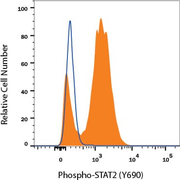
Detection of STAT2 in Daudi Human Cell Line by Flow Cytometry. Daudi human Burkitt's lymphoma cell line untreated (open histogram) or treated with 500 U/mL Recombinant Human IFN-aA (11100-1, filled histogram) for 20 minutes was stained with Rabbit Anti-Human Phospho-STAT2 (Y690) Monoclonal Antibody (Catalog # MAB2890), followed by Fluorescein-conjugated Anti-Rabbit IgG Secondary Antibody (F0112). To facilitate intracellular staining, cells were fixed with Flow Cytometry Fixation Buffer (FC004) and permeabilized with 90% methanol.
 View Larger
Detection of Human Phospho-STAT2 (Y690) by Simple WesternTM. Simple Western lane view shows lysates of Daudi human Burkitt's lymphoma cell line untreated (-) or treated (+) with 500 U/mL Recombinant Human IFN-aA (11100-1) for 20 minutes, loaded at 0.2 mg/mL. A specific band was detected for Phospho-STAT2 (Y690) at approximately 111 kDa (as indicated) using 0.5 µg/mL of Rabbit Anti-Human Phospho-STAT2 (Y690) Monoclonal Antibody (Catalog # MAB2890). This experiment was conducted under reducing conditions and using the 12-230 kDa separation system.
 View Larger
Detection of Human STAT2 by Western Blot Induction of type I IFN signaling by viral and nonviral stimuli is inhibited in G2/M-arrested cells. Suit2 cells were treated for 25 h with the vehicle, paclitaxel, colchicine, or ruxolitinib at 500 nM. Cells were then treated with the vehicle (untreated), TransIT reagent (0.5%, vol/vol), poly(I:C) at 10 μg/ml plus TransIT reagent, IFN-alpha at 5,000 U/ml, or VSV at an MOI of 30 based on titration on BHK-21 cells. VSV was aspirated 1 h later, and medium was added to infected wells. Cells remained in treatment for a total of 4 h, after which total protein was isolated. Western blot results for STAT1 and -2 proteins and their phosphorylated forms are shown in addition to VSV proteins. GAPDH was used to confirm that protein loading was the same across the gel. Protein names and protein sizes in kilodaltons are indicated on the left and right, respectively. Image collected and cropped by CiteAb from the following publication (https://pubmed.ncbi.nlm.nih.gov/30487274), licensed under a CC-BY license. Not internally tested by R&D Systems.
Reconstitution Calculator
Preparation and Storage
- 12 months from date of receipt, -20 to -70 °C as supplied.
- 1 month, 2 to 8 °C under sterile conditions after reconstitution.
- 6 months, -20 to -70 °C under sterile conditions after reconstitution.
Background: STAT2
STAT2 (signal transducer and activator of transcription 2) is a 113 kDa member of the STAT family of cytoplasmic transcription factors. STAT members generally mediate cytokine, growth factor and hormone receptor signal transduction. STAT2 is associated with type I ( alpha - and beta -) interferon signaling. All STATs contain an N‑terminal oligomerization domain, a DNA-binding domain, and an SH2-association region. STAT2 is phosphorylated at Y690 by receptor-associated Janus kinases (JAKs) leading to STAT2 dimerization and subsequent translocation to the nucleus to activate gene transcription.
Product Datasheets
Citations for Human Phospho-STAT2 (Y690) Antibody
R&D Systems personnel manually curate a database that contains references using R&D Systems products. The data collected includes not only links to publications in PubMed, but also provides information about sample types, species, and experimental conditions.
3
Citations: Showing 1 - 3
Filter your results:
Filter by:
-
Differential Responses of Human Dendritic Cells to Live or Inactivated Staphylococcus aureus: Impact on Cytokine Production and T Helper Expansion
Authors: Melania Cruciani, Silvia Sandini, Marilena P. Etna, Elena Giacomini, Romina Camilli, Martina Severa et al.
Frontiers in Immunology
-
Cell Cycle Arrest in G 2 /M Phase Enhances Replication of Interferon-Sensitive Cytoplasmic RNA Viruses via Inhibition of Antiviral Gene Expression
Authors: Christian Bressy, Gaith N. Droby, Bryant D. Maldonado, Nury Steuerwald, Valery Z. Grdzelishvili
Journal of Virology
-
Breaking resistance of pancreatic cancer cells to an attenuated vesicular stomatitis virus through a novel activity of IKK inhibitor TPCA-1
Authors: Marcela Cataldi, Nirav R. Shah, Sébastien A. Felt, Valery Z. Grdzelishvili
Virology
FAQs
No product specific FAQs exist for this product, however you may
View all Antibody FAQsReviews for Human Phospho-STAT2 (Y690) Antibody
There are currently no reviews for this product. Be the first to review Human Phospho-STAT2 (Y690) Antibody and earn rewards!
Have you used Human Phospho-STAT2 (Y690) Antibody?
Submit a review and receive an Amazon gift card.
$25/€18/£15/$25CAN/¥75 Yuan/¥2500 Yen for a review with an image
$10/€7/£6/$10 CAD/¥70 Yuan/¥1110 Yen for a review without an image


