Human R-Spondin 2 Antibody Summary
Met1-Gly205
Accession # Q6UXX9
Applications
Please Note: Optimal dilutions should be determined by each laboratory for each application. General Protocols are available in the Technical Information section on our website.
Scientific Data
 View Larger
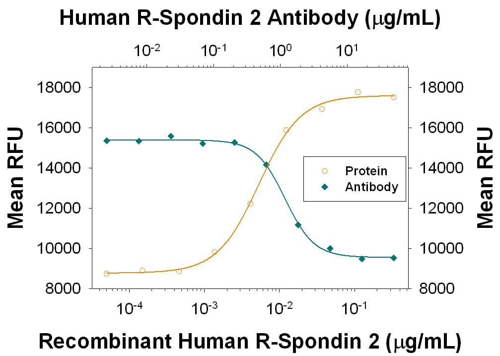
Β-Catenin Response Induced by R‑Spondin 2 and Neutral-ization by Human R‑Spondin 2 Antibody. Recombinant Human R-Spondin 2 (Catalog # 3266-RS) co-stimulates beta -catenin response in the HEK293T human embryonic kidney cell line in a dose-dependent manner (orange line). beta -Catenin response elicited by Recombinant Human R-Spondin 2 (30 ng/mL) is neutralized (green line) by increasing concentrations of Rat Anti-Human R-Spondin 2 Monoclonal Antibody (Catalog # MAB3266) in the presence of 5 ng/mL of Recombinant Mouse Wnt-3a (Catalog # 1324-WN). The ND50 is typically 0.4-2 µg/ml.
Preparation and Storage
- 12 months from date of receipt, -20 to -70 °C as supplied.
- 1 month, 2 to 8 °C under sterile conditions after reconstitution.
- 6 months, -20 to -70 °C under sterile conditions after reconstitution.
Background: R-Spondin 2
Human Roof plate-specific Spondin 2 isoform 1 (R-Spondin 2, RSPO-2), also known as cysteine-rich and single thrombospondin domain containing protein 2 (Cristin 2), is a 33 kDa secreted protein that belongs to the R-Spondin family. The four known human R-Spondins regulate beta -catenin signaling and overlap in expression and function (1‑3). Like other R-Spondins, R-Spondin 2 contains two adjacent cysteine-rich furin-like domains (aa 90-134) followed by a thrombospondin (TSP-1) motif (aa 144‑204) and a C-terminal region rich in basic residues (aa 207-243). The basic region appears to mediate cell surface retention but not to influence function (1). R‑Spondin 2 contains one potential N-glycosylation site. Of the three reported splice isoforms of human R-Spondin 2, isoform 2 lacks residues 1-67 of isoform 1, while isoform 3 has a glycine substitution for residues 32-95 of isoform 1. Human R-Spondin 2 is expressed in organs of endodermal origin in adults, including intestine and lung, and is downregulated in tumors of these tissues (1). In the embryonic mouse, R-Spondin 2 is expressed most highly in the hippocampus and in developing teeth and bones (4). Studies in Xenopus and mouse embryos indicate that R-Spondin 2 is an extracellular activator of Wnt/ beta -catenin signaling and is required for myogenesis (1). Mouse R-Spondin proteins bind both LRP-6 and Frizzled-8 but do not appear to form a ternary complex (3). R-Spondin 2 over-expression in Xenopus also blocks signaling of TGF-beta ligands, activin, nodal and BMP-4 (1). Human R-Spondin 2 is highly conserved across species, sharing 97-98% aa identity with mouse, rat, canine, bovine, and opossum R-Spondin 2 and 86% aa identity with Xenopus R-Spondin 2 within aa 22-205. Mature R-Spondin 2 shares ~40% aa identity with R-Spondin 1, R-Spondin 3, and R-Spondin 4.
- Kazanskaya, O. et al. (2004) Dev. Cell 7:525.
- Kim, K-A. et al. (2006) Cell Cycle 5:23.
- Nam, J-S. et al. (2006) J. Biol. Chem. 281:13247.
- Nam, J-S. et al. (2007) Gene Expr. 281:13247.
Product Datasheets
FAQs
No product specific FAQs exist for this product, however you may
View all Antibody FAQsReviews for Human R-Spondin 2 Antibody
There are currently no reviews for this product. Be the first to review Human R-Spondin 2 Antibody and earn rewards!
Have you used Human R-Spondin 2 Antibody?
Submit a review and receive an Amazon gift card.
$25/€18/£15/$25CAN/¥75 Yuan/¥2500 Yen for a review with an image
$10/€7/£6/$10 CAD/¥70 Yuan/¥1110 Yen for a review without an image


