Human RB1 Antibody Summary
Lys240-Asn406
Accession # P06400
Customers also Viewed
Applications
Please Note: Optimal dilutions should be determined by each laboratory for each application. General Protocols are available in the Technical Information section on our website.
Scientific Data
 View Larger
Detection of Human RB1 by Western Blot. Western blot shows lysates of Jurkat human acute T cell leukemia cell line, Daudi human Burkitt's lymphoma cell line, and Raji human Burkitt's lymphoma cell line. PVDF Membrane was probed with 0.1 µg/mL of Human RB1 Monoclonal Antibody (Catalog # MAB6495) followed by HRP-conjugated Anti-Mouse IgG Secondary Antibody (Catalog # HAF007). A specific band was detected for RB1 at approximately 120 kDa (as indicated). This experiment was conducted under reducing conditions and using Immunoblot Buffer Group 1.
 View Larger
RB1 in MCF‑7 Human Cell Line. RB1 was detected in immersion fixed MCF-7 human breast cancer cell line using Human RB1 Monoclonal Antibody (Catalog # MAB6495) at 10 µg/mL for 3 hours at room temperature. Cells were stained using the NorthernLights™ 557-conjugated Anti-Mouse IgG Secondary Antibody (red, upper panel; Catalog # NL007) and counterstained with DAPI (blue, lower panel). Specific staining was localized to nuclei. View our protocol for Fluorescent ICC Staining of Cells on Coverslips.
 View Larger
Detection of Human RB1 by Simple WesternTM. Simple Western lane view shows lysates of Jurkat human acute T cell leukemia cell line, loaded at 0.2 mg/mL. A specific band was detected for RB1 at approximately 120 kDa (as indicated) using 1 µg/mL of Mouse Anti-Human RB1 Monoclonal Antibody (Catalog # MAB6495). This experiment was conducted under reducing conditions and using the 12-230 kDa separation system.
 View Larger
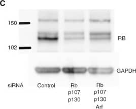
Detection of Human RB1 by Western Blot Simultaneous knockdown of multiple genes in primary human islets following electroporation. (A) Graph shows RT-qPCR results of islets electroporated with non-targeting control siRNA or siRNA targeting RB and p130 (L-003299). (B) Graph shows RT-qPCR results of islets electroporated with non-targeted control siRNA or siRNAs targeting the indicated gene products. The housekeeping gene GAPDH was used as the control. Representative experiment shown from 3 separate experiments. Error bars indicate standard deviation. (C) Western blot for RB protein in human islet lysates after electroporation of control or targeting siRNA. GAPDH was used as a loading control. RB appears as a characteristic doublet representing the hypo and hyperphosphorylated protein. Representative experiment shown from 3 different experiments. Image collected and cropped by CiteAb from the following open publication (https://pubmed.ncbi.nlm.nih.gov/25305068), licensed under a CC-BY license. Not internally tested by R&D Systems.
Preparation and Storage
- 12 months from date of receipt, -20 to -70 °C as supplied.
- 1 month, 2 to 8 °C under sterile conditions after reconstitution.
- 6 months, -20 to -70 °C under sterile conditions after reconstitution.
Background: RB1
Retinoblastoma 1 protein (RB-1; also retinoblastoma-associated protein, pp110, and p105-Rb) is a 110 kDa tumor suppressor gene and member of the retinoblastoma protein family. Human RB-1 is 928 amino acids in length. The protein contains a Pocket domain (aa 373-771), which is comprised of two other domains, domain A (aa 373-573) and domain B (aa 640-771), and a “spacer” (aa 580-639). The Pocket domain binds to threonine-phosphorylated domain C (aa 771-928), which thereby prevents interaction with heterodimeric E2F/DP transcription factor complexes. Human RB-1 is 90% aa identical to mouse RB-1. RB-1 is expressed in the retina. The underphosphorylated, active form of RB-1 interacts with E2F1 and represses its transcription activity, leading to cell cycle arrest. Defects in RB-1 lead to the childhood cancer retinoblastoma.
Product Datasheets
Citations for Human RB1 Antibody
R&D Systems personnel manually curate a database that contains references using R&D Systems products. The data collected includes not only links to publications in PubMed, but also provides information about sample types, species, and experimental conditions.
6
Citations: Showing 1 - 6
Filter your results:
Filter by:
-
Divergent transcriptional and transforming properties of PAX3-FOXO1 and PAX7-FOXO1 paralogs
Authors: Manceau L, Richard Albert J, Lollini PL et al.
PLOS Genetics
-
Divergent transcriptional and transforming properties of PAX3-FOXO1 and PAX7-FOXO1 paralogs
Authors: Manceau L, Richard Albert J, Lollini PL et al.
PLOS Genetics
-
Effect of miR‑215 on the Expression of Tumor Suppressor Gene Rb1 in Retinoblastoma Cell Lines
Authors: Liqin SHAO, Zhangxing SHENG, Yuefeng ZHU, Jianchao LI, Rufa MENG
Iranian Journal of Public Health
-
Ubiquitin C‐terminal hydrolase‐L1 has prognostic relevance and is a therapeutic target for high‐grade neuroendocrine lung cancers
Authors: Yoshihisa Shimada, Yujin Kudo, Sachio Maehara, Jun Matsubayashi, Yoichi Otaki, Naohiro Kajiwara et al.
Cancer Science
-
Simultaneous silencing of multiple RB and p53 pathway members induces cell cycle reentry in intact human pancreatic islets
Authors: Stanley Tamaki, Christopher Nye, Euan Slorach, David Scharp, Helen M Blau, Phyllis E Whiteley et al.
BMC Biotechnology
-
Simultaneous silencing of multiple RB and p53 pathway members induces cell cycle reentry in intact human pancreatic islets
Authors: Stanley Tamaki, Christopher Nye, Euan Slorach, David Scharp, Helen M Blau, Phyllis E Whiteley et al.
BMC Biotechnology
Species: Human
Sample Types: Cell Lysates
Applications: Western Blot
FAQs
No product specific FAQs exist for this product, however you may
View all Antibody FAQsReviews for Human RB1 Antibody
There are currently no reviews for this product. Be the first to review Human RB1 Antibody and earn rewards!
Have you used Human RB1 Antibody?
Submit a review and receive an Amazon gift card.
$25/€18/£15/$25CAN/¥75 Yuan/¥2500 Yen for a review with an image
$10/€7/£6/$10 CAD/¥70 Yuan/¥1110 Yen for a review without an image


