Human RPA2 Antibody Summary
Leu141-Glu270
Accession # P15927
Applications
Please Note: Optimal dilutions should be determined by each laboratory for each application. General Protocols are available in the Technical Information section on our website.
Scientific Data
 View Larger
Detection of Human RPA2 by Western Blot. Western blot shows lysates of HeLa human cervical epithelial carcinoma cell line, Saos-2 human osteosarcoma cell line, SH-SY5Y human neuroblastoma cell line, and human tonsil tissue. PVDF membrane was probed with 1 µg/mL of Goat Anti-Human RPA2 Antigen Affinity-purified Polyclonal Antibody (Catalog # AF5937) followed by HRP-conjugated Anti-Goat IgG Secondary Antibody (HAF019). A specific band was detected for RPA2 at approximately 36 kDa (as indicated). This experiment was conducted under reducing conditions and using Immunoblot Buffer Group 8.
 View Larger
Detection of Human RPA2 by Simple WesternTM. Simple Western lane view shows lysates of Jurkat human acute T cell leukemia cell line, U2OS human osteosarcoma cell line, MCF‑7 human breast cancer cell line, and HEK293T human embryonic kidney cell line, loaded at 0.2 mg/mL. A specific band was detected for RPA2 at approximately 38 kDa (as indicated) using 25 µg/mL of Goat Anti-Human RPA2 Antigen Affinity-purified Polyclonal Antibody (Catalog # AF5937). This experiment was conducted under reducing conditions and using the 12-230 kDa separation system.
 View Larger
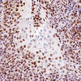
Detection of RPA2 in Human Tonsil. RPA2 was detected in immersion fixed paraffin-embedded sections of Human Tonsil using Goat Anti-Human RPA2 Antigen Affinity-purified Polyclonal Antibody (Catalog # AF5937) at 5 µg/mL for 1 hour at room temperature followed by incubation with the Anti-Goat IgG VisUCyte™ HRP Polymer Antibody (Catalog # VC004). Before incubation with the primary antibody, tissue was subjected to heat-induced epitope retrieval using VisUCyte Antigen Retrieval Reagent-Basic (Catalog # VCTS021). Tissue was stained using DAB (brown) and counterstained with hematoxylin (blue). Specific staining was localized to cell nuclei. View our protocol for IHC Staining with VisUCyte HRP Polymer Detection Reagents.
Preparation and Storage
- 12 months from date of receipt, -20 to -70 °C as supplied.
- 1 month, 2 to 8 °C under sterile conditions after reconstitution.
- 6 months, -20 to -70 °C under sterile conditions after reconstitution.
Background: RPA2
RPA2 (replication protein A 32 kDa subunit; also RFA2 and RPA p34) is a 32 kDa DNA‑binding protein that constitutess one of three subunits comprising the PRA heterotrimer complex. In conjunction with 70 kDa RPA1 and 14 kDa RPA3, RPA2 participates in DNA replication, recombination and repair. Human RPA2 is 270 amino acids (aa) in length. It contains a Gly/Ser‑rich N‑terminus (aa 1‑33), a DNA‑binding domain (aa 43‑171) and a protein‑interaction C‑terminus (aa 187‑270). Phosphorylation of the N‑terminus on Ser4/8/23/29/33, plus Thr21, regulates RPA complex interactions with DNA repair and replication complexes. There are multiple splice variants. Three contain N‑terminal extensions: one shows an 88 aa insertion after Ser4, another shows a 12 aa substitution for aa 1‑4, and a third shows a four aa insertion after Ser4. There is also a deletion of aa 93‑98, and a potential truncation after Gln175. Over aa 141‑270, human RPA2 shares 83% aa identity with mouse RPA2.
Product Datasheets
FAQs
No product specific FAQs exist for this product, however you may
View all Antibody FAQsReviews for Human RPA2 Antibody
There are currently no reviews for this product. Be the first to review Human RPA2 Antibody and earn rewards!
Have you used Human RPA2 Antibody?
Submit a review and receive an Amazon gift card.
$25/€18/£15/$25CAN/¥75 Yuan/¥2500 Yen for a review with an image
$10/€7/£6/$10 CAD/¥70 Yuan/¥1110 Yen for a review without an image
