Mouse CD55/DAF Antibody Summary
Asp35-Pro359
Accession # Q61475
Applications
Please Note: Optimal dilutions should be determined by each laboratory for each application. General Protocols are available in the Technical Information section on our website.
Scientific Data
 View Larger
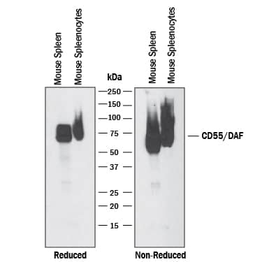
Detection of Mouse CD55/DAF by Western Blot. Western blot shows lysates of mouse spleen tissue and mouse splenocytes. PVDF membrane was probed with 1 µg/mL of Sheep Anti-Mouse CD55/DAF Antigen Affinity-purified Polyclonal Antibody (Catalog # AF5376) followed by HRP-conjugated Anti-Sheep IgG Secondary Antibody (Catalog # HAF016). A specific band was detected for CD55/DAF at approximately 75 kDa (as indicated). This experiment was conducted under reducing and non-reducing conditions and using Immunoblot Buffer Group 1.
 View Larger
Detection of CD55/DAF in Mouse Splenocytes by Flow Cytometry. Mouse splenocytes were stained with Sheep Anti-Mouse CD55/DAF Antigen Affinity-purified Polyclonal Antibody (Catalog # AF5376, filled histogram) or control antibody (Catalog # 5-001-A, open histogram), followed by NorthernLights™ 637-conjugated Anti-Sheep IgG Secondary Antibody (Catalog # NL011).
 View Larger
Detection of Mouse CD55/DAF by Western Blot Glimepiride releases CD14 from RAW 264 cells. (A) The amounts of CD14 in RAW 264 cells treated for one hour with control medium (□) or glimepiride (■) as shown. Values are means ± SD, from triplicate experiments performed 4 times, n = 12. (B) The amounts of CD14 in supernatants from RAW 264 cells treated for one hour with control medium (□) or glimepiride as shown (■). Values are means ± SD, from triplicate experiments performed 4 times, n = 12. (C) Immunoblots showing the amounts of CD14, PrPC, CD55 and caveolin in extracts from RAW 264 cells treated for 1 hour with control medium (i) or 5 μM glimepiride (ii). (D) The amounts of CD14 in cells (□) or supernatants (■) from microglial cells treated for 1 hour with control medium, 5 μM glimepiride or 5 μM glipizide. Values are mean units CD14 ± SD, from triplicate experiments performed 3 times, n = 9. *Cellular CD14 significantly less than control cells. **supernatant CD14 significantly greater than control supernatants. (E) Blot showing the amounts of CD14 in supernatants from microglial cells treated with concentrations of glimepiride as shown for one hour. Image collected and cropped by CiteAb from the following publication (https://pubmed.ncbi.nlm.nih.gov/24952384), licensed under a CC-BY license. Not internally tested by R&D Systems.
Reconstitution Calculator
Preparation and Storage
- 12 months from date of receipt, -20 to -70 °C as supplied.
- 1 month, 2 to 8 °C under sterile conditions after reconstitution.
- 6 months, -20 to -70 °C under sterile conditions after reconstitution.
Background: CD55/DAF
CD55 (Decay-accelarating factor/DAF) is a glycoprotein member of the RCA family of molecules. It is found on blood cells, epithelium and endothelium and serves both as a receptor for CD97 and a negative regulator of the C3 convertases, C4b2a and C3bBb. Mature mouse CD55 is the product of two genes that arose by duplication. There is a 55-60 kDa, 356 amino acid (aa), GPI-linked form that is ubiquitously expressed. This molecule contains four SUSHI domains (aa 35-285), a Ser/Thr-rich region (aa 288-362) and a GPI-anchor at Gly362. There is also a 50 kDa, 379 aa, type I transmembrane form that is testis-associated. It shows the same domain architecture and is 93% aa identical to the GPI-form. At least four GPI gene isoforms exist. They diverge after Ile285 and show deletions and substitutions. Over aa 35-359, mouse CD55 is 66% and 50% aa identical to rat and human CD55, respectively.
Product Datasheets
Citation for Mouse CD55/DAF Antibody
R&D Systems personnel manually curate a database that contains references using R&D Systems products. The data collected includes not only links to publications in PubMed, but also provides information about sample types, species, and experimental conditions.
1 Citation: Showing 1 - 1
-
Glimepiride reduces CD14 expression and cytokine secretion from macrophages.
Authors: Ingham V, Williams A, Bate C
J Neuroinflammation, 2014-06-21;11(0):115.
Species: Mouse
Sample Types: Cell Lysates
Applications: Western Blot
FAQs
No product specific FAQs exist for this product, however you may
View all Antibody FAQsReviews for Mouse CD55/DAF Antibody
There are currently no reviews for this product. Be the first to review Mouse CD55/DAF Antibody and earn rewards!
Have you used Mouse CD55/DAF Antibody?
Submit a review and receive an Amazon gift card.
$25/€18/£15/$25CAN/¥75 Yuan/¥2500 Yen for a review with an image
$10/€7/£6/$10 CAD/¥70 Yuan/¥1110 Yen for a review without an image
