Mouse CD83 Antibody Summary
Met22-Ala134
Accession # O88324
Applications
Please Note: Optimal dilutions should be determined by each laboratory for each application. General Protocols are available in the Technical Information section on our website.
Scientific Data
 View Larger
CD83 in Mouse Splenocytes. CD83 was detected in immersion fixed mouse splenocytes using 10 µg/mL Goat Anti-Mouse CD83 Antigen Affinity-purified Polyclonal Antibody (Catalog # AF1437) for 3 hours at room temperature. Cells were stained with the NorthernLights™ 557-conjugated Anti-Goat IgG Secondary Antibody (red; Catalog # NL001) and counterstained with DAPI (blue). View our protocol for Fluorescent ICC Staining of Non-adherent Cells.
 View Larger
CD83 in Mouse Dendritic Cells. CD83 was detected in immersion fixed LPS-stimulated mouse dendritic cells using Goat Anti-Mouse CD83 Antigen Affinity-purified Polyclonal Antibody (Catalog # AF1437) at 10 µg/mL for 3 hours at room temperature. Cells were stained using the NorthernLights™ 557-conjugated Anti-Goat IgG Secondary Antibody (red; Catalog # NL001) and counterstained with DAPI (blue). View our protocol for Fluorescent ICC Staining of Non-adherent Cells.
Reconstitution Calculator
Preparation and Storage
- 12 months from date of receipt, -20 to -70 °C as supplied.
- 1 month, 2 to 8 °C under sterile conditions after reconstitution.
- 6 months, -20 to -70 °C under sterile conditions after reconstitution.
Background: CD83
Mouse CD83 is a 30‑35 kDa member of the Siglec (or sialic-acid-binding immunoglobulin-like lectin) family of transmembrane proteins (1, 2, 3). CD83 is synthesized as a type I transmembrane glycoprotein that contains a 114 amino acid (aa) extracellular region, a 22 aa transmembrane segment, and a 39 aa cytoplasmic domain. It contains one V type Ig-like domain in the extracellular region with no inhibitory cytoplasmic motif(s). In the extracellular region, mouse and human CD83 are 66% aa identical (1, 2, 4). Relative to mouse, human CD83 has an 11 aa insertion in its extracellular domain and is expressed as a 45‑55 kDa protein (1, 4, 5, 6). No alternate splice variants have been reported for mouse. In human, however, one soluble splice form has been reported and proteolytic processing is suggested to generate a second circulating isoform (6, 7). Notably, although soluble CD83 has the potential to exist as either a monomer or disulfide-linked dimer, both show immunosuppressive activity (4, 8, 9). Membrane CD83, by contrast, is immunostimulatory (10). CD83 is a primary marker for dendritic cells (3, 5, 6). It is also found on B cells (6, 11), neutrophils (12), monocytes and macrophages (13). Except for dendritic cells, CD83 expression is often transient. CD83 binds to sialic acids on monocytes (3). The function of CD83 is only now becoming clear. As noted, membrane-immobilized CD83 appears to promote T cell proliferation, particularly of CD8+ cytotoxic T cells (14). On monocytes, CD83 may also drive monocytes into a fibrocyte phenotype (14). And a lack of membrane-expressed CD83 leads to an unusual IL-4/IL-10 producing CD4+ T cell phenotype (15).
- Berchtold, S. et. al. (1999) FEBS Lett. 461:211.
- Fujimoto, Y. and T.F. Tedder (2006) J. Med. Dent. Sci. 53:85.
- Scholler, N. et. al. (2001) J. Immunol. 166:3865.
- Lechmann, M. et al. (2005) Biochem. Biophys. Res. Commun. 329:132.
- Zhou, L-J. et. al. (1992) J. Immunol. 149:735.
- Hock, B.D. et al. (2001) Int. Immunol. 13:959.
- Dudziak, D. et al. (2005) J. Immunol. 174:6672.
- Kotzor, N. et al. (2004) Immunobiology 209:129.
- Zinser, E. et al. (2006) Immunobiology 21:449.
- Hirano, N. et al. (2006) Blood 107:1528.
- Cramer, S.O. et al. (2000) Int. Immunol. 12:1347.
- Yamashiro, S. et al. (2000) Blood 96:3958.
- Cao, W. et al. (2005) Biochem. J. 385:85.
- Scholler, N. et al. (2002) J. Immunol. 168:2599.
- Garcia-Martinez, L.F. et al. (2004) J. Immunol. 173:2995.
Product Datasheets
Citations for Mouse CD83 Antibody
R&D Systems personnel manually curate a database that contains references using R&D Systems products. The data collected includes not only links to publications in PubMed, but also provides information about sample types, species, and experimental conditions.
4
Citations: Showing 1 - 4
Filter your results:
Filter by:
-
CD83 mediates the inhibitory effect of the S1PR1 agonist CYM5442 on LPS-induced M1 polarization of macrophages through the ERK-STAT-1 signaling pathway
Authors: Luo, M;Zhang, W;Yang, J;Du, X;Wang, X;Xu, G;Tang, H;Wang, Z;Zhong, X;Feng, J;Ma, N;
International immunopharmacology
Species: Mouse
Sample Types: Cell Lysates
Applications: Western Blot -
Dendritic cell CD83 homotypic interactions regulate inflammation and promote mucosal homeostasis.
Authors: Bates J, Flanagan K, Mo L, Ota N, Ding J, Ho S, Liu S, Roose-Girma M, Warming S, Diehl L
Mucosal Immunol, 2014-09-10;8(2):414-28.
Species: Mouse
Sample Types: Cell Culture Supernates
Applications: ELISA Development (Capture) -
Inactivated Sendai virus particles eradicate tumors by inducing immune responses through blocking regulatory T cells.
Authors: Kurooka M, Kaneda Y
Cancer Res., 2007-01-01;67(1):227-36.
Species: Mouse
Sample Types: Whole Cells
Applications: Flow Cytometry -
CD83 expressed by macrophages is an important immune checkpoint molecule for the resolution of inflammation
Authors: Katrin Peckert-Maier, Pia Langguth, Astrid Strack, Lena Stich, Petra Mühl-Zürbes, Christine Kuhnt et al.
Frontiers in Immunology
FAQs
No product specific FAQs exist for this product, however you may
View all Antibody FAQsReviews for Mouse CD83 Antibody
Average Rating: 5 (Based on 1 Review)
Have you used Mouse CD83 Antibody?
Submit a review and receive an Amazon gift card.
$25/€18/£15/$25CAN/¥75 Yuan/¥2500 Yen for a review with an image
$10/€7/£6/$10 CAD/¥70 Yuan/¥1110 Yen for a review without an image
Filter by:
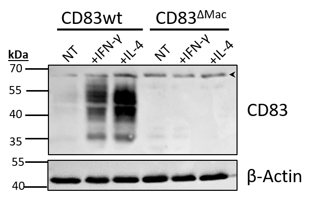
Bone marrow-derived macrophages from wt or conditional KO mice were stimulated either with IFN-y or IL-4 for 16 h. Whole cell lysates were prepared and 50 µg of protein was used for SDS-PAGE (16%) and western blotting (primary anti-CD83 1:500; secondary anti-goat-HRP 1:7500).
Specific signal only in WT-macrophages, no signal in KO cells.
One unspecific band at approx. 65 kDa (see arrow-head).


