Mouse CHL-1/L1CAM-2 Antibody Summary
Ala25-Gln1043 (Leu227-Gln242 del, Ala243Ser)
Accession # BAC30699
Applications
Please Note: Optimal dilutions should be determined by each laboratory for each application. General Protocols are available in the Technical Information section on our website.
Scientific Data
 View Larger
Detection of Mouse CHL‑1/L1CAM‑2 by Western Blot. Western blot shows lysates of mouse brain stem tissue. PVDF membrane was probed with 0.25 µg/mL of Goat Anti-Mouse CHL-1/L1CAM-2 Antigen Affinity-purified Polyclonal Antibody (Catalog # AF2147) followed by HRP-conjugated Anti-Goat IgG Secondary Antibody (Catalog # HAF019). A specific band was detected for CHL-1/L1CAM-2 at approximately 200 kDa (as indicated). This experiment was conducted under reducing conditions and using Immunoblot Buffer Group 1.
 View Larger
Detection of Mouse CHL‑1/L1CAM‑2 by Simple WesternTM. Simple Western lane view shows lysates of mouse brain (cortex) tissue and mouse brain stem tissue, loaded at 0.2 mg/mL. A specific band was detected for CHL-1/L1CAM-2 at approximately 196-201 kDa (as indicated) using 2.5 µg/mL of Goat Anti-Mouse CHL-1/L1CAM-2 Antigen Affinity-purified Polyclonal Antibody (Catalog # AF2147) followed by 1:50 dilution of HRP-conjugated Anti-Goat IgG Secondary Antibody (Catalog # HAF109). This experiment was conducted under reducing conditions and using the 12-230 kDa separation system.
 View Larger
Detection of Mouse CHL-1/L1CAM-2 by Western Blot BACE1-mediated processing of APP and CHL1 is reduced in cortex of aged BACE1 cKO mice following tamoxifen treatment. Cortex homogenates from TAM- or VEH-treated mice were resolved by SDS-PAGE for Western blot analysis of APP and CHL1 processing. Homogenates from aged-matched BACE+/− and BACE1−/− were also loaded as control samples. APP-FL, pC99 and pC89 were normalized to GAPDH (MAB374) while CHL1-FL and CHL1-NTF were normalized to beta -tubulin (JDR.3B8). Protein amount was normalized to protein levels in control mice injected with vehicle (set at 1). Representative blots of (a) APP-FL (C1/6.1), (b) APP-CTFs (C1/6.1) and (c) CHL1. (d) Densitometry analysis of protein expression. APP processing was reduced in TAM-treated mice as demonstrated by the accumulation of APP-FL (C1/6.1), and reduced levels of the beta CTFs pC99 and pC89. beta CTFs were clearly identified because missing in the BACE1−/− sample. CHL1-FL (AF2147) levels were increased and CHL1-NTF levels were significantly reduced. Furthermore, the CHL1-NTF/CHL1-FL ratio was significantly decreased in TAM-treated mice demonstrating reduced BACE1 processing (VEH n = 7; TAM n = 7). (e) Quantification of A beta x-40 was performed by MSD immunoassay on cortex homogenates and expressed as pMol/g of cortex. The decrease of levels of A beta x-40 in TAM-treated mice was comparable to the one observed in samples collected from young TAM-treated mice (~50% decrease) (VEH n = 7; TAM n = 7). Results were plotted as Mean ± SEM, *p < 0.05; **p < 0.005; ***p < 0.001; ****p < 0.0001; n.s. = not significant, Student’s t test. Image collected and cropped by CiteAb from the following publication (https://pubmed.ncbi.nlm.nih.gov/31882662), licensed under a CC-BY license. Not internally tested by R&D Systems.
 View Larger
Detection of Mouse CHL-1/L1CAM-2 by Western Blot Axon guidance defects were absent in hippocampus mossy fibers of aged BACE1 cKO mice following partial BACE1 deletion. (a) Coronal sections collected from aged mice were stained with anti-synaptoporin (SPO) antibody (green) and DAPI (blue). Scale bar 50 μm. (b) Quantification of IPB length showed no alteration in TAM-treated mice compared to controls. IPB length was normalized on the length of the CA3 stratum lucidum (VEH n = 8; TAM n = 7, 3 to 4 sections per mouse). (c) Representative microscopy images showing reduced BACE1 (D10E5) expression in the hippocampus of TAM-treated mice. BACE1 signal was totally absent in BACE−/− mice, used as control to evaluate the amount of background in the staining. Scale bar 200 μm. Hippocampus full homogenates from TAM- or VEH-treated mice were resolved by SDS-PAGE for analysis of APP processing and fractionated (soluble and membrane fractions) for the analysis of SEZ6 and CHL1 processing. Homogenates from aged-matched BACE+/− and BACE1−/− were loaded as control samples. Representative blots of (d) APP-FL (C1/6.1) and APP- CTFs (C1/6.1), (e) fractionation blots of sAPP beta (BAWT), SEZ6 (14E5) and CHL1 (AF2147). (f) Densitometry analysis of protein expression. APP processing was reduced in TAM-treated mice as demonstrated by the accumulation of APP-FL (C1/6.1), and reduced levels of the beta CTFs pC99 and pC89, and sAPP beta. beta CTFs and sAPP beta were identified because missing in the BACE1−/− sample. SEZ6 processing was decreased in TAM-treated mice with accumulation of the full length and decreased levels of the ectodomain (SEZ6-NTF) as well as decreased SEZ6-NTF/SEZ6FL ratio. Processing of CHL1 was also impaired as showed by increased of CHL1-FL levels, while CHL1-NTF was not altered. CHL1-NTF/CHL1-FL ratio was significantly decreased. APP-FL, CTFs, SEZ6-NTF and CHL1-NTF were normalized to GAPDH (MAB374), SEZ6-FL and CHL1-FL were normalized to Calnexin (610523) (VEH n = 5; TAM n = 5). (g) A beta x-40 was quantified from hippocampus homogenates by MSD immunoassay. TAM-treated group displayed a significant reduction of A beta x-40 levels (~50% decrease) compared to control (VEH n = 7; TAM n = 7). Results were plotted as Mean ± SEM, **p < 0.005; ***p < 0.001; n.s. = not significant, Student’s t test. DG: dentate gyrus, IPB: infrapyramidal bundle, slu: stratum lucidum, MB: main bundle. Image collected and cropped by CiteAb from the following publication (https://pubmed.ncbi.nlm.nih.gov/31882662), licensed under a CC-BY license. Not internally tested by R&D Systems.
 View Larger
Detection of Mouse CHL-1/L1CAM-2 by Western Blot BACE1-mediated processing of APP and CHL1 is reduced in cortex of young BACE1 cKO mice following tamoxifen treatment. Cortex homogenates from TAM- or VEH-treated mice were resolved by SDS-PAGE for Western blot analysis of APP and CHL1 processing. Homogenates from aged-matched BACE+/− and BACE1−/− were also loaded as control samples. Representative blots of (a) APP-full length (APP-FL) (C1/6.1), (b) APP-Carboxy Terminal Fragments (CTFs) (C1/6.1) and (c) CHL1. (d) Densitometry analysis of protein expression. Protein amount was normalized to protein levels in control mice (set at 1). APP-FL, pC99 and pC89 were normalized to GAPDH (MAB374) while CHL1-FL and CHL1-NTF were normalized to beta -tubulin (JDR.3B8). APP processing was reduced in TAM-treated mice as demonstrated by the accumulation of APP-FL (C1/6.1), and reduced levels of the beta CTFs pC99 and pC89. beta CTFs were clearly identified because missing in the BACE1−/− sample. CHL1-FL (AF2147) levels were increased while CHL1-N Terminal Fragment (CHL1-NTF) levels were not affected in cortex of TAM-treated mice. However, the CHL1-NTF/CHL1-FL ratio was significantly decreased in TAM-treated mice demonstrating reduced BACE1 processing (VEH n = 8; TAM n = 8). (e) A beta x-40 was quantified from brain homogenates by ELISA (VEH n = 8; TAM n = 8). Levels of A beta x-40 expressed as pMol/g of cortex were significantly reduced in TAM-treated mice (~50% decrease). Results were plotted as Mean ± SEM, ***p < 0.001; ****p < 0.0001; n.s. = not significant, Student’s t test. Image collected and cropped by CiteAb from the following publication (https://pubmed.ncbi.nlm.nih.gov/31882662), licensed under a CC-BY license. Not internally tested by R&D Systems.
 View Larger
Detection of Mouse Mouse CHL-1/L1CAM-2 Antibody by Western Blot Quantitative proteomics in Rab35 cKO P0 hippocampus.a Volcano plot of the TMT-based quantitative proteomes identifying the dysregulated proteins in Rab35 cKO hippocampus in comparison with the control hippocampus (n = 5 mice per genotype). b Number of proteins identified as significantly dysregulated and as either membrane traffic-related or neuronal migration-related. c, d Western blot analysis of control and Rab35 cKO P0 hippocampi using anti-contactin-2, anti-CHL1, and anti-actin antibodies. e, f Quantification of contactin-2 (c) and CHL1 (d) protein levels in control and Rab35 cKO P0 hippocampi. Band intensities of the indicated proteins were normalized to those of actin (n = 9 mice per genotype). Unpaired Student’s t-test; e, p = 0.0153; f, p = 0.0095. g, h Levels of contactin-2 (g) and CHL1 (h) were quantified by targeted MS using the PRM method (n = 5 mice per genotype). Unpaired Student’s t-test; gp = 0.0053; hp = 0.0229. i Western blot analysis of control and Rab35 cKO P0 hippocampus using anti-N-cadherin and anti-actin antibodies. j Quantification of N-cadherin protein levels in the control and Rab35 cKO P0 hippocampus (n = 9 mice per genotypes). Unpaired Student’s t-test, p = 0.9020. k Representative images of DIV 2 hippocampal primary neurons stained for contactin-2 (green), rhodamine-phalloidin (magenta) and DAPI (blue). Scale bar, 20 μm. l Quantification of contactin-2 intensity at the somatic plasma membrane in control (n = 4) and Rab35-deficient (n = 4) cells. Thirty neurons from four different cultures per genotype were analyzed. Mann–Whitney U-test, p = 0.0286. Data represent the mean ± SEM; n.s. not significant (p > 0.05); *p < 0.05; **p < 0.01. Image collected and cropped by CiteAb from the following publication (https://pubmed.ncbi.nlm.nih.gov/37085665), licensed under a CC-BY license. Not internally tested by R&D Systems.
Reconstitution Calculator
Preparation and Storage
- 12 months from date of receipt, -20 to -70 °C as supplied.
- 1 month, 2 to 8 °C under sterile conditions after reconstitution.
- 6 months, -20 to -70 °C under sterile conditions after reconstitution.
Background: CHL-1/L1CAM-2
Close homolog of L1 (CHL-1), also known as cell adhesion L1-like (CALL) and L1 cell adhesion molecule 2 (L1CAM-2), belongs to the L1 subfamily of immunoglobulin (Ig) superfamily cell adhesion molecules, which also includes L1, neurofascin and NgCAM-related cell adhesion molecule (NrCAM) (1‑3). These molecules are type I transmembrane proteins that have 6 Ig-like domains and 4‑5 fibronectin type III-like (FNIII) domains in their extracellular regions. They also share a highly conserved cytoplasmic region of approximately 110 amino acid (aa) residues containing an ankyrin-binding site. CHL-1 is expressed as a highly glycosylated 185 kDa transmembrane protein by subpopulations of neurons and glia of the central and peripheral nervous system (4, 5). Ectodomain shedding via the metalloprotease-disintegrin ADAM8 releases 165 kDa and 125 kDa soluble CHL-1 fragments, which can diffuse away to function at distant sites (6). CHL-1 is not capable of homotypic interactions, but an extracellular binding partner of CHL-1 has not been identified (4). Human CHL1 has been mapped to chromosome 3p26 and is a candidate gene for 3p- syndrome characterized by mental impairment (7). A missense CHL1 polymorphism associated increased risk of schizophrenia, has also been reported (8). The functional importance of CHL-1 in the nervous system is also evident in CHL-1 deficient mice, which display behavioral abnormalities and show misguided axons within the hippocampus and olfactory tract (9). Enhanced ectodomain-shedding of CHL-1 is also observed in Wobbler mice, the neurodegenerative mutant mice (6). In vitro, soluble or substrate-coated CHL-1 promotes neurite outgrowth and neuronal survival of both cerebellar and hippocampal neurons. Cell surface CHL-1 interacts with integrins in cis to potentiate integrin-dependent cell migration toward extracellular matrix proteins (10). For this enhanced cell motility, CHL-1 linkage to the actin cytoskeleton via interaction between ankyrin and the CHL-1 cytoplasmic region is required.
- Moos, M. et al. (1988) Nature 334:701.
- Holm, J. et al. (1996) Eur. J. Neusci. 8:1613.
- Wei, M. et al. (1998) Hum. Genet. 103:355.
- Hillenbrand, R. et al. (1999) Eur. J. Neurosci. 11:813.
- Liu, Q. et al. (2000) J. Neurosci. 20:7682.
- Naus, S. et al. (2004) J. Biol. Chem. 279:16083.
- Angeloni, D. et al. (1999) Am. J. Med. Genet. 86:482.
- Sakurai, K. et al. (2002) Mol. Psychiatry 7:412.
- Montag-Sallaz, M. et al. (2002) Mol. Cell. Biol. 22:7967.
- Buhusi, M. et al. (2003) J. Biol. Chem. 278(27):25024.
Product Datasheets
Citations for Mouse CHL-1/L1CAM-2 Antibody
R&D Systems personnel manually curate a database that contains references using R&D Systems products. The data collected includes not only links to publications in PubMed, but also provides information about sample types, species, and experimental conditions.
25
Citations: Showing 1 - 10
Filter your results:
Filter by:
-
RAB35 is required for murine hippocampal development and functions by regulating neuronal cell distribution
Authors: Ikuko Maejima, Taichi Hara, Satoshi Tsukamoto, Hiroyuki Koizumi, Takeshi Kawauchi, Tomoko Akuzawa et al.
Communications Biology
-
Reduced secretion of neuronal growth regulator 1 contributes to impaired adipose-neuronal crosstalk in obesity
Authors: E Duregotti, CM Reumiller, U Mayr, M Hasman, LE Schmidt, SA Burnap, K Theofilato, J Barallobre, A Beran, M Grandoch, A Viviano, M Jahangiri, M Mayr
Nature Communications, 2022-11-25;13(1):7269.
Species: Mouse
Sample Types: Tissue Homogenates, Whole Tissue
Applications: IHC, Western Blot -
Proteomic and functional analyses of the periodic membrane skeleton in neurons
Authors: R Zhou, B Han, R Nowak, Y Lu, E Heller, C Xia, AH Chishti, VM Fowler, X Zhuang
Nature Communications, 2022-06-09;13(1):3196.
Species: Mouse
Sample Types: Whole Cells
Applications: ICC -
Interplay in neural functions of cell adhesion molecule close homolog of L1 (CHL1) and Programmed Cell Death 6 (PDCD6)
Authors: Gabriele Loers, Thomas Theis, Helen Baixia Hao, Ralf Kleene, Sanjana Arsha, Nina Samuel et al.
FASEB BioAdvances
-
Spatiotemporal processing of neural cell adhesion molecules 1 and 2 by BACE1 in vivo
Authors: W Kim, H Watanabe, S Lomoio, G Tesco
The Journal of Biological Chemistry, 2021-02-03;0(0):100372.
Species: Mouse
Sample Types: Tissue Homogenates
Applications: Western Blot -
BACE1 partial deletion induces synaptic plasticity deficit in adult mice
Authors: S Lombardo, M Chiacchiar, A Tarr, W Kim, T Cao, G Sigal, TW Rosahl, W Xia, PG Haydon, ME Kennedy, G Tesco
Sci Rep, 2019-12-27;9(1):19877.
Species: Mouse
Sample Types: Tissue Homogenates
Applications: Western Blot -
Proximity proteomics identifies cancer cell membrane cis ‐molecular complex as a potential cancer target
Authors: Norihiro Kotani, Arisa Yamaguchi, Tomoko Ohnishi, Ryusuke Kuwahara, Takanari Nakano, Yuka Nakano et al.
Cancer Science
-
Axonal growth of midbrain dopamine neurons is modulated by the cell adhesion molecule ALCAM through trans-heterophilic interaction with L1Cam, ChL1 and Semaphorin
Authors: CR Bye, V Rytova, WF Alsanie, CL Parish, LH Thompson
J. Neurosci., 2019-07-12;0(0):.
Species: Mouse
Sample Types: Whole Cells, Whole Tissue
Applications: Cell Culture, IHC -
Close Homolog of L1 Regulates Dendritic Spine Density in the Mouse Cerebral Cortex through Semaphorin 3B
Authors: V Mohan, SD Wade, CS Sullivan, MR Kasten, C Sweetman, R Stewart, Y Truong, M Schachner, PB Manis, PF Maness
J. Neurosci., 2019-06-10;0(0):.
Species: Mouse
Sample Types: Tissue Homogenates, Whole Cells, Whole Tissue
Applications: ICC, IHC, Immunoprecipitation, Western Blot -
Axo-axonic Innervation of Neocortical Pyramidal Neurons by GABAergic Chandelier Cells Requires AnkyrinG-Associated L1CAM
Authors: Yilin Tai, Nicholas B. Gallo, Minghui Wang, Jia-Ray Yu, Linda Van Aelst
Neuron
-
BACE1 elevation engendered by GGA3 deletion increases ?-amyloid pathology in association with APP elevation and decreased CHL1 processing in 5XFAD mice
Authors: W Kim, L Ma, S Lomoio, R Willen, S Lombardo, J Dong, PG Haydon, G Tesco
Mol Neurodegener, 2018-02-02;13(1):6.
Species: Mouse
Sample Types: Tissue Homogenates
Applications: Western Blot -
Proteolytic cleavage of transmembrane cell adhesion molecule L1 by extracellular matrix molecule Reelin is important for mouse brain development
Authors: David Lutz, Ahmed Sharaf, Dagmar Drexler, Hardeep Kataria, Gerrit Wolters-Eisfeld, Bianka Brunne et al.
Scientific Reports
-
A beta -dependent reduction of NCAM2-mediated synaptic adhesion contributes to synapse loss in Alzheimer’s disease
Authors: Iryna Leshchyns’ka, Heng Tai Liew, Claire Shepherd, Glenda M. Halliday, Claire H. Stevens, Yazi D. Ke et al.
Nature Communications
-
Antagonistic Effects of BACE1 and APH1B-gamma -Secretase Control Axonal Guidance by Regulating Growth Cone Collapse
Authors: Soraia Barão, Annette Gärtner, Eduardo Leyva-Díaz, Galina Demyanenko, Sebastian Munck, Tine Vanhoutvin et al.
Cell Reports
-
Transcriptome analysis reveals transmembrane targets on transplantable midbrain dopamine progenitors.
Authors: Bye C, Jonsson M, Bjorklund A, Parish C, Thompson L
Proc Natl Acad Sci U S A, 2015-03-09;112(15):E1946-55.
Species: Mouse, Rat
Sample Types: Tissue Homogenates, Whole Tissue
Applications: Flow Cytometry, IHC -
An aberrant sugar modification of BACE1 blocks its lysosomal targeting in Alzheimer's disease.
Authors: Kizuka Y, Kitazume S, Fujinawa R, Saito T, Iwata N, Saido T, Nakano M, Yamaguchi Y, Hashimoto Y, Staufenbiel M, Hatsuta H, Murayama S, Manya H, Endo T, Taniguchi N
EMBO Mol Med, 2015-02-01;7(2):175-89.
Species: Mouse
Sample Types: Tissue Homogenates
Applications: Western Blot -
Endothelial VEGF Sculpts Cortical Cytoarchitecture
Authors: Suyan Li, Katharina Haigh, Jody J. Haigh, Anju Vasudevan
The Journal of Neuroscience
-
beta-Site amyloid precursor protein (APP)-cleaving enzyme 1 (BACE1)-deficient mice exhibit a close homolog of L1 (CHL1) loss-of-function phenotype involving axon guidance defects.
Authors: Hitt B, Riordan S, Kukreja L, Eimer W, Rajapaksha T, Vassar R
J Biol Chem, 2012-09-17;287(46):38408-25.
Species: Mouse
Sample Types: Whole Tissue
Applications: IHC-Fr -
Neuronal cadherin (NCAD) increases sensory neurite formation and outgrowth on astrocytes
Authors: Toby A. Ferguson, Steven S. Scherer
Neuroscience Letters
-
Secretome protein enrichment identifies physiological BACE1 protease substrates in neurons.
Authors: Kuhn PH, Koroniak K, Hogl S, Colombo A, Zeitschel U, Willem M, Volbracht C, Schepers U, Imhof A, Hoffmeister A, Haass C, Rossner S, Brase S, Lichtenthaler SF
EMBO J., 2012-06-22;31(14):3157-68.
Species: Mouse
Sample Types: Cell Lysates
Applications: Western Blot -
EphB regulates L1 phosphorylation during retinocollicular mapping.
Mol. Cell. Neurosci., 2012-05-08;50(2):201-10.
Species: Mouse
Sample Types: Cell Lysates
Applications: Immunoprecipitation, Western Blot -
CHL1 negatively regulates the proliferation and neuronal differentiation of neural progenitor cells through activation of the ERK1/2 MAPK pathway.
Authors: Huang X, Zhu LL, Zhao T
Mol. Cell. Neurosci., 2010-10-08;46(1):296-307.
Species: Mouse
Sample Types: Whole Tissue
Applications: IHC-Fr -
Phosphatidylinositol 3-kinase/protein kinase Cdelta activation induces close homolog of adhesion molecule L1 (CHL1) expression in cultured astrocytes.
Authors: Wu J, Wrathall JR, Schachner M
Glia, 2010-02-01;58(3):315-28.
Species: Mouse
Sample Types: Cell Lysates, Whole Cells
Applications: ICC, Neutralization, Western Blot -
CHL1 promotes Sema3A-induced growth cone collapse and neurite elaboration through a motif required for recruitment of ERM proteins to the plasma membrane.
Authors: Schlatter MC, Buhusi M, Wright AG, Maness PF
J. Neurochem., 2007-11-06;104(3):731-44.
Species: Mouse
Sample Types: Whole Cells
Applications: ICC -
Tau and other proteins found in Alzheimer's disease spinal fluid are linked to retromer-mediated endosomal traffic in mice and humans.
Authors: Simoes S, Neufeld J. L, et al.
Sci Transl Med
FAQs
No product specific FAQs exist for this product, however you may
View all Antibody FAQsReviews for Mouse CHL-1/L1CAM-2 Antibody
Average Rating: 5 (Based on 3 Reviews)
Have you used Mouse CHL-1/L1CAM-2 Antibody?
Submit a review and receive an Amazon gift card.
$25/€18/£15/$25CAN/¥75 Yuan/¥2500 Yen for a review with an image
$10/€7/£6/$10 CAD/¥70 Yuan/¥1110 Yen for a review without an image
Filter by:
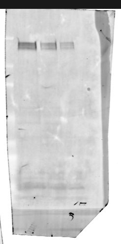
80, 40 and 20ug of total mouse brain homogenate was separated by SDS-PAGE and transfered to PVDF membrane
Block: 1% BSA, 1% FSG PBS-T 1h RT
Primary: 1:1000, 1% BSA, 1% FSG PBS-T O/N 4oC
Secondary: 1:5000,1% BSA, 1% FSG PBS-T 2h RT
(Secondary - DaG 800, Licor)


