Mouse Coagulation Factor III/Tissue Factor Antibody
Mouse Coagulation Factor III/Tissue Factor Antibody Summary
Ala29-Glu251
Accession # P20352
*Small pack size (-SP) is supplied either lyophilized or as a 0.2 µm filtered solution in PBS.
Applications
Please Note: Optimal dilutions should be determined by each laboratory for each application. General Protocols are available in the Technical Information section on our website.
Scientific Data
 View Larger
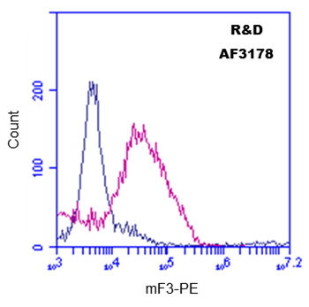
Detection of Coagulation Factor III/Tissue Factor in Raw 264.7 cells treated with 1 µg/mL LPS overnight by Flow Cytometry Raw 264.7 cells treated with 1 µg/mL LPS overnight were stained with Goat Anti-Mouse Coagulation Factor III/Tissue Factor Antigen Affinity-purified Polyclonal Antibody (Catalog # AF3178, filled histogram) or isotype control antibody (Catalog # AB-108-C, open histogram) followed by Phycoerythrin-conjugated Anti-Goat IgG Secondary Antibody (Catalog # F0107). View our protocol for Staining Membrane-associated Proteins.
Reconstitution Calculator
Preparation and Storage
- 12 months from date of receipt, -20 to -70 °C as supplied.
- 1 month, 2 to 8 °C under sterile conditions after reconstitution.
- 6 months, -20 to -70 °C under sterile conditions after reconstitution.
Background: Coagulation Factor III/Tissue Factor
Coagulation Factor III/Tissue Factor (TF), also known as thromboplastin and CD142, is an integral membrane protein found in a variety of cell types. It functions as a protein cofactor/receptor of Coagulation Factor VII, which is synthesized in the liver and circulated in the plasma (1). Upon binding of TF, the inactive factor VII is rapidly converted into activated VIIa. The resulting 1:1 complex of VIIa and TF initiates the coagulation pathway and has also important coagulation-independent functions such as angiognesis (2). Synthesized as a 294 amino acid precursor, mouse TF consists of a signal peptide (residues 1-28) and the mature chain (residues 29-294). As a type I membrane protein, it contains a transmembrane region (residues 252-274) and a cytoplasmic tail (residues 275-294) (3, 4). The purified rmTF corresponds to the ectodomain (residues 29-251) and is potent in activating thermolysin-processed rmCoagulation Factor VII (R&D Systems, Catalog # 3305-SE) under the conditions described in the Activity Assay Protocol.
- Morrissey, J.H. (2004) in Handbook of Proteolytic Enzymes. Barrett, A.J. et al. (ed) Academic Press, San Diego, p. 1659.
- Versteeg, H.H. et al. (2003) Carcinogenesis 24:1009.
- Ranganathan, G. et al. (1991) J. Biol. Chem. 266:496.
- Hartzell, S. (1989) Mol. Cell. Biol. 9:2567.
Product Datasheets
Citations for Mouse Coagulation Factor III/Tissue Factor Antibody
R&D Systems personnel manually curate a database that contains references using R&D Systems products. The data collected includes not only links to publications in PubMed, but also provides information about sample types, species, and experimental conditions.
17
Citations: Showing 1 - 10
Filter your results:
Filter by:
-
Evaluation of different commercial antibodies for their ability to detect human and mouse tissue factor by western blotting
Authors: Rosell A, Moser B, Hisada Y et al.
Res Pract Thromb Haemost
-
Aging impairs cold-induced beige adipogenesis and adipocyte metabolic reprogramming
Authors: CD Holman, AP Sakers, RP Calhoun, L Cheng, EC Fein, C Jacobs, L Tsai, ED Rosen, P Seale
bioRxiv : the preprint server for biology, 2023-03-23;0(0):.
Species: Mouse
Sample Types: Whole Cells
Applications: Flow Cytometry -
Hypoxia induced up-regulation of tissue factor is mediated through extracellular RNA activated Toll-like receptor 3-activated protein 1 signalling
Authors: Saumya Bhagat, Indranil Biswas, Rehan Ahmed, Gausal A. Khan
Blood Cells, Molecules, and Diseases
-
SENP3 in monocytes/macrophages up-regulates tissue factor and mediates lipopolysaccharide-induced acute lung injury by enhancing JNK phosphorylation
Authors: X Chen, Y Lao, J Yi, J Yang, S He, Y Chen
J. Cell. Mol. Med., 2020-03-31;0(0):.
Species: Mouse
Sample Types: Tissue Lysate
Applications: Western Blot -
Adipocytes express tissue factor and FVII and are procoagulant in a TF/FVIIa-dependent manner
Authors: Desirée Edén, Grigorios Panagiotou, Dariush Mokhtari, Jan W. Eriksson, Mikael Åberg, Agneta Siegbahn
Upsala Journal of Medical Sciences
-
Protease-activated receptor 2 protects against VEGF inhibitor-induced glomerular endothelial and podocyte injury
Authors: Y Oe, T Fushima, E Sato, A Sekimoto, K Kisu, H Sato, J Sugawara, S Ito, N Takahashi
Sci Rep, 2019-02-27;9(1):2986.
Species: Mouse
Sample Types: Whole Tissue
Applications: IHC-P -
Enzymatic lipid oxidation by eosinophils propagates coagulation, hemostasis, and thrombotic disease
Authors: Stefan Uderhardt, Jochen A. Ackermann, Tobias Fillep, Victoria J. Hammond, Johann Willeit, Peter Santer et al.
Journal of Experimental Medicine
-
Leukocyte integrin Mac-1 regulates thrombosis via interaction with platelet GPIb alpha
Authors: Yunmei Wang, Huiyun Gao, Can Shi, Paul W. Erhardt, Alexander Pavlovsky, Dmitry A. Soloviev et al.
Nature Communications
-
The Myosin II Inhibitor, Blebbistatin, Ameliorates FeCl3-induced Arterial Thrombosis via the GSK3?-NF-?B Pathway
Authors: Y Zhang, L Li, Y Zhao, H Han, Y Hu, D Liang, B Yu, J Kou
Int. J. Biol. Sci., 2017-05-15;13(5):630-639.
Species: Mouse
Sample Types: Whole Tissue
Applications: IHC -
Protective and detrimental effects of neuroectodermal cell–derived tissue factor in mouse models of stroke
Authors: Shaobin Wang, Brandi Reeves, Erica M. Sparkenbaugh, Janice Russell, Zbigniew Soltys, Hua Zhang et al.
JCI Insight
-
Hepatocyte tissue factor contributes to the hypercoagulable state in a mouse model of chronic liver injury
Authors: Pierre-Emmanuel Rautou, Kohei Tatsumi, Silvio Antoniak, A. Phillip Owens, Erica Sparkenbaugh, Lori A. Holle et al.
Journal of Hepatology
-
Inflammation drives thrombosis after Salmonella infection via CLEC-2 on platelets
Authors: Jessica R. Hitchcock, Charlotte N. Cook, Saeeda Bobat, Ewan A. Ross, Adriana Flores-Langarica, Kate L. Lowe et al.
Journal of Clinical Investigation
-
Tissue factor/factor VIIa signalling promotes cytokine-induced beta cell death and impairs glucose-stimulated insulin secretion from human pancreatic islets
Authors: Desirée Edén, Agneta Siegbahn, Dariush Mokhtari
Diabetologia
-
Regulation of Alveolar Procoagulant Activity and Permeability in Direct Acute Lung Injury by Lung Epithelial Tissue Factor
Authors: Ciara M. Shaver, Brandon S. Grove, Nathan D. Putz, Jennifer K. Clune, William E. Lawson, Robert H. Carnahan et al.
American Journal of Respiratory Cell and Molecular Biology
-
Tissue factor-targeted lidamycin inhibits growth and metastasis of colon carcinoma
Authors: QING ZHANG, XIUJUN LIU, CAIHONG LI, DONGSHENG LIAO, ZHIGANG OUYANG, JUNNIAN ZHENG et al.
Oncology Letters
-
Mechanical stretch inhibits lipopolysaccharide-induced keratinocyte-derived chemokine and tissue factor expression while increasing procoagulant activity in murine lung epithelial cells.
Authors: Sebag S, Bastarache J, Ware L
J Biol Chem, 2013-01-29;288(11):7875-84.
Species: Mouse
Sample Types: Cell Lysates
Applications: Western Blot -
Elevated tissue factor expression contributes to exacerbated diabetic nephropathy in mice lacking eNOS fed a high fat diet.
Authors: Li F, Wang CH, Wang JG, Thai T, Boysen G, Xu L, Turner AL, Wolberg AS, Mackman N, Maeda N, Takahashi N
J. Thromb. Haemost., 2010-10-01;8(10):2122-32.
Species: Mouse
Sample Types: In Vivo
Applications: Neutralization
FAQs
No product specific FAQs exist for this product, however you may
View all Antibody FAQsReviews for Mouse Coagulation Factor III/Tissue Factor Antibody
Average Rating: 5 (Based on 4 Reviews)
Have you used Mouse Coagulation Factor III/Tissue Factor Antibody?
Submit a review and receive an Amazon gift card.
$25/€18/£15/$25CAN/¥75 Yuan/¥2500 Yen for a review with an image
$10/€7/£6/$10 CAD/¥70 Yuan/¥1110 Yen for a review without an image
Filter by:
This protein might be found in high levels in some conditions/tissues. If you calibrate a good concentration you will have a nice band around 50 KDa
Used as detection antibody after labeling with Sulfo-Tag according to manufacturer's protocol (Meso Scale Diagnostics LLC)
Detection range - 8-30,000 pg/ml
Detection of Coagulation Factor III in Mouse mononuclear cells using Goat anti-Mouse F3 antibody (#AF3178, pink) at 2.5 ug/10^6 cells, or Isotype Control IgG (blue), followed by PE-conjugated anti-Goat secondary antibody.


