Mouse Midkine Antibody Summary
Gly89-Asp140
Accession # P12025
Applications
Please Note: Optimal dilutions should be determined by each laboratory for each application. General Protocols are available in the Technical Information section on our website.
Scientific Data
 View Larger
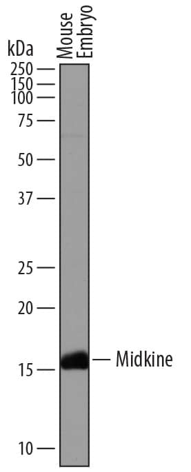
Detection of Mouse Midkine by Western Blot. Western blot shows lysates of mouse embryo tissue. PVDF membrane was probed with 1 µg/mL of Sheep Anti-Mouse Midkine Antigen Affinity-purified Polyclonal Antibody (Catalog # AF7769) followed by HRP-conjugated Anti-Sheep IgG Secondary Antibody (Catalog # HAF016). A specific band was detected for Midkine at approximately 15-17 kDa (as indicated). This experiment was conducted under reducing conditions and using Immunoblot Buffer Group 1.
 View Larger
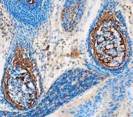
Midkine in Mouse Embryo. Midkine was detected in immersion fixed paraffin-embedded sections of mouse embryo (13 d.p.c.) using Sheep Anti-Mouse Midkine Antigen Affinity-purified Polyclonal Antibody (Catalog # AF7769) at 5 µg/mL overnight at 4 °C. Tissue was stained using the Anti-Sheep HRP-DAB Cell & Tissue Staining Kit (brown; Catalog # CTS019) and counterstained with hematoxylin (blue). Specific staining was localized to cartilage primordium in ribs. View our protocol for Chromogenic IHC Staining of Paraffin-embedded Tissue Sections.
 View Larger
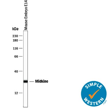
Detection of Human and Mouse Midkine by Simple WesternTM. Simple Western lane view shows lysates of mouse embryo tissue, loaded at 0.2 mg/mL. A specific band was detected for Midkine at approximately 26 kDa (as indicated) using 20 µg/mL of Sheep Anti-Mouse Midkine Antigen Affinity-purified Polyclonal Antibody (Catalog # AF7769) followed by 1:50 dilution of HRP-conjugated Anti-Goat IgG Secondary Antibody (Catalog # HAF109). This experiment was conducted under reducing conditions and using the 12-230 kDa separation system.
Preparation and Storage
- 12 months from date of receipt, -20 to -70 °C as supplied.
- 1 month, 2 to 8 °C under sterile conditions after reconstitution.
- 6 months, -20 to -70 °C under sterile conditions after reconstitution.
Background: Midkine
Midkine (MK; also Retinoid acid-induced differentiation facor) is a secreted heparin-binding member of the very small pleiotrophin family of proteins. Although its predicted MW is 13 kDa, it runs anomalously at 15-17 kDa in SDS-PAGE. MK is strongly expressed in the embryo, but is known to be secreted in the adult by endothelium, preadipocytes, proximal renal tubular epithelium, and CD4+ T cells. MK has multiple activities, including the inhibition of regulatory T cell production, the promotion of adipocyte formation, and the induction of chemokine production by smooth muscle, the clustering of Ach receptors on myoblasts, and the migration of embryonic neurons plus neutrophils and macrophages. It has multiple receptors, including heparin and chondroitin sulfate, LRP-1, nucleolin, ALK, and PTP-zeta. Mature mouse midkine is 118 amino acids (aa) in length (aa 23-140). It possesses two distinct domains, an N-terminal domain spanning aa 23-71, and a C-terminal domain that encompasses aa 81-140. The C-terminal domain is further divided into two basic amino acid clusters that bind heparin. There are two splice variants reported for mouse midkine. One shows a deletion of aa 39-103, while another shows a deletion of aa 80-133. Midkine will form a covalent, crosslinked homodimer through the action of tissue type II transglutaminase. Full-length mature mouse MK (aa 23-140) shares 97% and 86% aa sequence with full-length rat and human MK, respectively..
Product Datasheets
Citation for Mouse Midkine Antibody
R&D Systems personnel manually curate a database that contains references using R&D Systems products. The data collected includes not only links to publications in PubMed, but also provides information about sample types, species, and experimental conditions.
1 Citation: Showing 1 - 1
-
Validation and in vivo characterization of research antibodies for Moesin, CD44, Midkine, and sFRP-1.
Authors: Doolen, S;Ayoubi, R;Laflamme, C;Betarbet, R;Zoeller, E;Williams, S;Fu, H;Levey, A;Rizzo, S;
F1000Research
Species: Mouse
Sample Types: Cell Lysates, Tissue Homogenates
Applications: Western Blot
FAQs
No product specific FAQs exist for this product, however you may
View all Antibody FAQsReviews for Mouse Midkine Antibody
There are currently no reviews for this product. Be the first to review Mouse Midkine Antibody and earn rewards!
Have you used Mouse Midkine Antibody?
Submit a review and receive an Amazon gift card.
$25/€18/£15/$25CAN/¥75 Yuan/¥2500 Yen for a review with an image
$10/€7/£6/$10 CAD/¥70 Yuan/¥1110 Yen for a review without an image

