Mouse MMR/CD206 Antibody Summary
Leu19-Ala1388
Accession # Q2HZ94
Applications
Please Note: Optimal dilutions should be determined by each laboratory for each application. General Protocols are available in the Technical Information section on our website.
Scientific Data
 View Larger
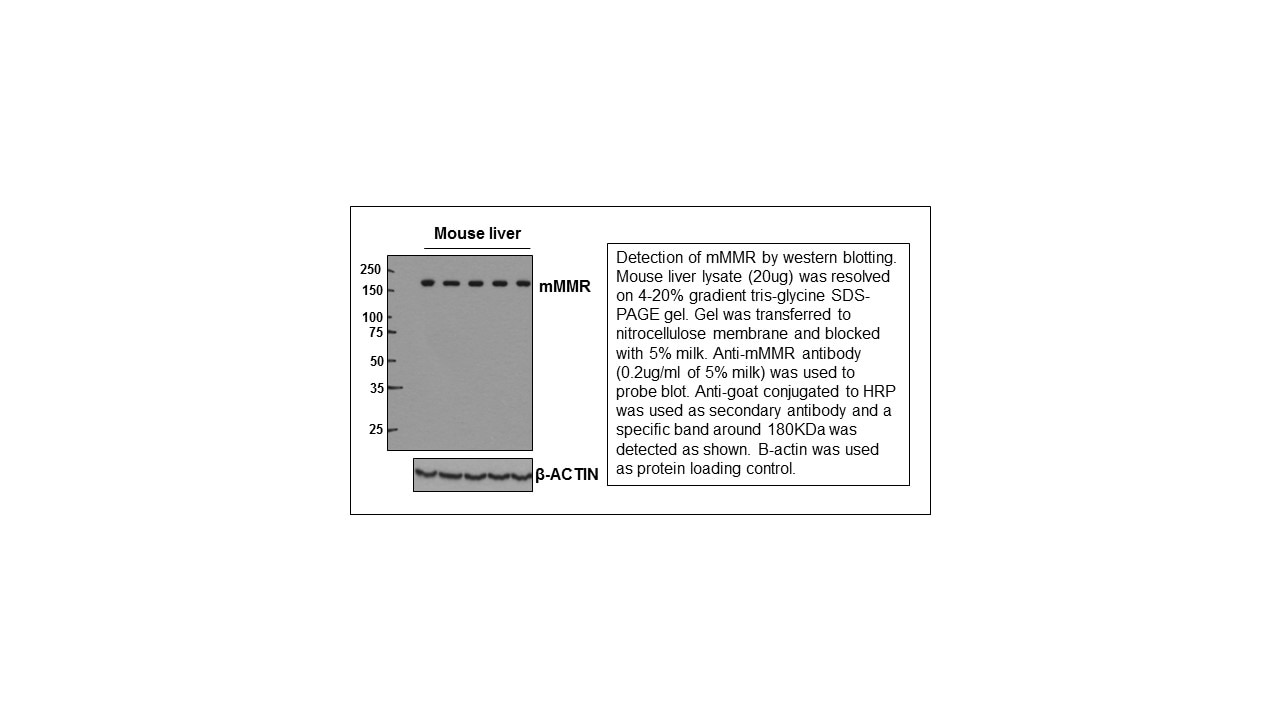
Detection of Mouse MMR/CD206 by Western Blot. Western blot shows lysates of mouse liver tissue. Gels were loaded with 12 µg, 6.5 µg, and 3 µg of tissue lysate. PVDF membrane was probed with 1 µg/mL of Goat Anti-Mouse MMR/CD206 Antigen Affinity-purified Polyclonal Antibody (Catalog # AF2535) followed by HRP-conjugated Anti-Goat IgG Secondary Antibody (Catalog # HAF019). A specific band was detected for MMR/CD206 at approximately 180 kDa (as indicated). This experiment was conducted under reducing conditions and using Immunoblot Buffer Group 1.
 View Larger
MMR/CD206 in Mouse Testis. MMR/CD206 was detected in perfusion fixed frozen sections of mouse testis using Goat Anti-Mouse MMR/CD206 Antigen Affinity-purified Polyclonal Antibody (Catalog # AF2535) at 5 µg/mL overnight at 4 °C. Tissue was stained using the Anti-Goat HRP-DAB Cell & Tissue Staining Kit (brown; Catalog # CTS008) and counterstained with hematoxylin (blue). Specific staining was localized to spermatocytes in testis. View our protocol for Chromogenic IHC Staining of Frozen Tissue Sections.
 View Larger
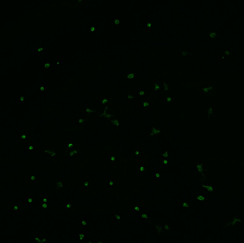
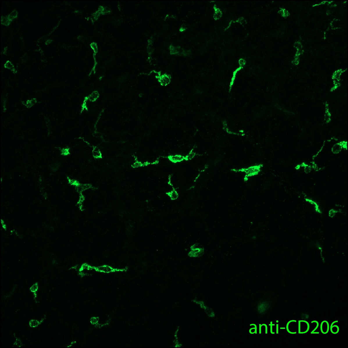
MMR/CD206 in Mouse Lung. MMR/CD206 was detected in perfusion fixed frozen sections of mouse lung using Goat Anti-Mouse MMR/CD206 Antigen Affinity-purified Polyclonal Antibody (Catalog # AF2535) at 25 µg/mL overnight at 4 °C. Tissue was stained using the NorthernLights™ 493-conjugated Anti-Goat IgG Secondary Antibody (green; Catalog # NL003) and counterstained with DAPI (blue). Specific staining was localized to cytoplasm of macrophages. View our protocol for Fluorescent IHC Staining of Frozen Tissue Sections.
 View Larger
Detection of Human MMR/CD206/Mannose Receptor by Immunocytochemistry/Immunofluorescence Cells of human meninges co-express LLEC markers. a–c DAB-IHC with single antibodies detects VEGFR3 (a), LYVE1 (b), and MRC1 (c) in the meninges of human post mortem brain showing no signs of neuropathology. These images are taken from a 38 year old male (sample P17/07, Table 1), and confirmed in n = 2 additional samples. P parenchyma. Scale = 150 µm (a); 40 µm (b); and 20 µm (c). d–f DAB-IHC with single antibodies detects VEGFR3 (b), LYVE1 (c), and MRC1 (d) in elderly human meninges (age: 89–92) with evidence of neuropathology and confirmed in n = 3 brains (Table 1). P, parenchyma. Scale = 20 µm. g–p IHC with fluorescent antibodies detects human meningeal cells that co-express MRC1 (h, m, yellow), LYVE1 (i, n, white), and VEGFR3 (j, o, green). Nuclei/RNA are labelled with DAPI (g, l, blue) and images are merged in (k, p). Scale = 10 µm Image collected and cropped by CiteAb from the following publication (https://pubmed.ncbi.nlm.nih.gov/31696318), licensed under a CC-BY license. Not internally tested by R&D Systems.
 View Larger
Detection of Mouse MMR/CD206/Mannose Receptor by Immunocytochemistry/Immunofluorescence M1 and M2 phenotype in spinal cord after intraplantar IL-1 beta. Wild-type (WT) and LysM-G protein–coupled receptor kinase (GRK)2+/− mice received an intraplantar injection of 1 ng IL-1 beta. At 15 hours after injection, spinal cord was collected, and frozen sections of (A) lumbar spinal cord (L2 to L5) and as control (B) thoracic spinal cord (T6 to T10) were stained for M1 (CD16/32) and M2 (CD206 and arginase-I) phenotypic markers. A representative example of M1 and M2 staining in the dorsal horn of one of the four mice per group is displayed. Scale bar indicates 20 μm. (C) Quantification of microglia/macrophages expressing M1 and M2 phenotypic markers in spinal cord from WT and LysM-GRK2+/− mice. Expression was quantified in approximately 10 to 15 dorsal horns of spinal cords per group (4 mice per group). The level of expression in the lumbar or thoracic area from control WT mice was set at 100%. Data are expressed as means ± SEM. **P < 0.01, ***P < 0.001. Image collected and cropped by CiteAb from the following publication (https://pubmed.ncbi.nlm.nih.gov/22731384), licensed under a CC-BY license. Not internally tested by R&D Systems.
 View Larger
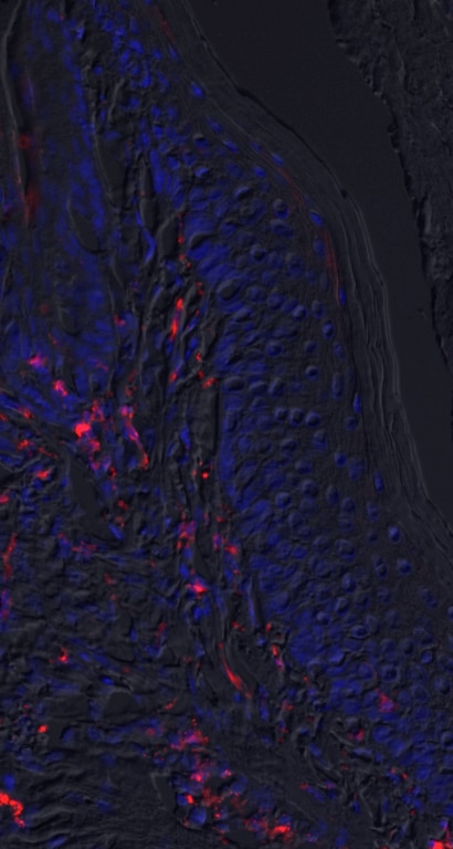
Detection of Mouse MMR/CD206/Mannose Receptor by Immunocytochemistry/Immunofluorescence Immunofluorescent staining for macrophage marker F4/80 in Acomys and Mus.(A–C) Bone-marrow-derived cells isolated from Acomys and stained for F4/80 (green). (A) unstimulated cells, (B) cells stimulated with IFN gamma and LPS, (C) cells stimulated with IL-4. (D–F) Bone-marrow-derived cells isolated from Mus and stained for F4/80 (green). (D) unstimulated cells, (E) cells stimulated with IFN gamma and LPS, and (F) cells stimulated with IL-4. Scale bar = 50 μm. (G) Acomys ear tissue at D15 after injury stained for F4/80 (green), CD206 (red) and DAPI (grey). (H) Mus ear tissue at D7 after injury stained for F4/80 (green), CD206 (red) and DAPI (grey). Scale bar = 50 μm.DOI:https://dx.doi.org/10.7554/eLife.24623.012 Image collected and cropped by CiteAb from the following publication (https://pubmed.ncbi.nlm.nih.gov/28508748), licensed under a CC-BY license. Not internally tested by R&D Systems.
 View Larger
Detection of Mouse MMR/CD206/Mannose Receptor by Immunohistochemistry Characterization of M2 BV2 cells induced by IL-4 and identification of sEVs derived from M2 BV2 cells. (A, B) Representative images of BV2 cells immunostained for Iba-1 (green), CD206 (red), and arginase (red). Cultured systems were treated with 0 or 20 ng/µL IL-4. Cell nuclei were counterstained with DAPI. Scale bar = 50 µm. (C) Western blotting analysis of CD206 and arginase expression in BV2 cells after 0 or 20 ng/µL IL-4 treatment. (D) Representative electron microscopy images showing the phenotype of M2-sEVs. Left image scale bar = 100 nm, right image scale bar = 50 nm. (F) NTA of M2-sEVs isolated by ultracentrifugation from M2 BV2 cells. Data represent the average size distribution profile of three samples and each purification normalized to the total nanoparticle concentrations. Data for each sample was derived from three different videos and analyses. (G) Western blotting analysis of TSG101 and CD63 levels in M2 BV2 cells and M2-sEVs. Image collected and cropped by CiteAb from the following publication (https://pubmed.ncbi.nlm.nih.gov/33391532), licensed under a CC-BY license. Not internally tested by R&D Systems.
 View Larger
Detection of Human MMR/CD206/Mannose Receptor by Immunocytochemistry/Immunofluorescence Cells of human meninges co-express LLEC markers. a–c DAB-IHC with single antibodies detects VEGFR3 (a), LYVE1 (b), and MRC1 (c) in the meninges of human post mortem brain showing no signs of neuropathology. These images are taken from a 38 year old male (sample P17/07, Table 1), and confirmed in n = 2 additional samples. P parenchyma. Scale = 150 µm (a); 40 µm (b); and 20 µm (c). d–f DAB-IHC with single antibodies detects VEGFR3 (b), LYVE1 (c), and MRC1 (d) in elderly human meninges (age: 89–92) with evidence of neuropathology and confirmed in n = 3 brains (Table 1). P, parenchyma. Scale = 20 µm. g–p IHC with fluorescent antibodies detects human meningeal cells that co-express MRC1 (h, m, yellow), LYVE1 (i, n, white), and VEGFR3 (j, o, green). Nuclei/RNA are labelled with DAPI (g, l, blue) and images are merged in (k, p). Scale = 10 µm Image collected and cropped by CiteAb from the following publication (https://pubmed.ncbi.nlm.nih.gov/31696318), licensed under a CC-BY license. Not internally tested by R&D Systems.
 View Larger
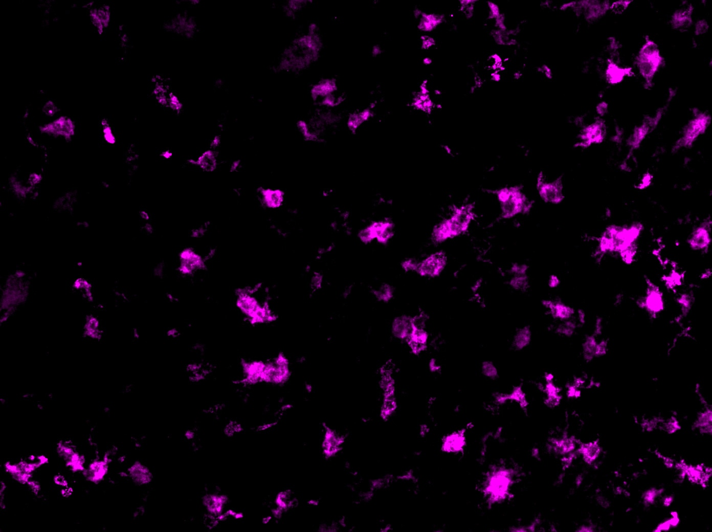
Detection of Mouse MMR/CD206/Mannose Receptor by Immunocytochemistry/Immunofluorescence Mouse LLECs take up A beta 1-40. a Schematic showing the site of dye and A beta 1-40 perfusion into the CSF via the cisterna magna (arrow) of a 2-month old mouse. The dotted line indicates the plane of section. A anterior, P posterior, D dorsal, V ventral. b Coronal brain section indicating the areas imaged. SF4 refers to area captured in Figure S4. c The percentage of each labelled cell type that internalized perfused A beta. Cells co-expressing VEGFR3 and LYVE1 take up A beta at a higher rate than MRC1, LYVE1 double-positive cells as well as MRC1-positive, LYVE1-negative cells (p ≤ 0.05, bootstrap). VEGFR3, LYVE1 counts, n = 2 brains (3 sections/brain). MRC1, LYVE1 counts, n = 3 brains (3 sections/brain). d–d′′′ Cells of the adult mouse meninges that co-express VEGFR3 (d, green) and LYVE1 (d′, white) internalize A beta 1-40 (d′′, cyan). Scale = 20 µm. e-e′′′) Cells of the adult mouse meninges that co-express VEGFR3 (e, green) and MRC1 (e′, white) internalize A beta 1-40 (e′′, cyan). Scale = 40 µm. f–f′′′) Cells of the adult mouse meninges that co-express MRC1 (f, magenta) and LYVE1 (f′, white) internalize A beta 1-40 (f′′, cyan). The walls of a blood vessel (white arrowhead, f′′) also accumulate A beta 1-40. Scale = 60 µm Image collected and cropped by CiteAb from the following publication (https://pubmed.ncbi.nlm.nih.gov/31696318), licensed under a CC-BY license. Not internally tested by R&D Systems.
 View Larger
Detection of Human MMR/CD206/Mannose Receptor by Immunocytochemistry/Immunofluorescence Cells of human meninges co-express LLEC markers. a–c DAB-IHC with single antibodies detects VEGFR3 (a), LYVE1 (b), and MRC1 (c) in the meninges of human post mortem brain showing no signs of neuropathology. These images are taken from a 38 year old male (sample P17/07, Table 1), and confirmed in n = 2 additional samples. P parenchyma. Scale = 150 µm (a); 40 µm (b); and 20 µm (c). d–f DAB-IHC with single antibodies detects VEGFR3 (b), LYVE1 (c), and MRC1 (d) in elderly human meninges (age: 89–92) with evidence of neuropathology and confirmed in n = 3 brains (Table 1). P, parenchyma. Scale = 20 µm. g–p IHC with fluorescent antibodies detects human meningeal cells that co-express MRC1 (h, m, yellow), LYVE1 (i, n, white), and VEGFR3 (j, o, green). Nuclei/RNA are labelled with DAPI (g, l, blue) and images are merged in (k, p). Scale = 10 µm Image collected and cropped by CiteAb from the following publication (https://pubmed.ncbi.nlm.nih.gov/31696318), licensed under a CC-BY license. Not internally tested by R&D Systems.
 View Larger
Detection of Mouse MMR/CD206/Mannose Receptor by Immunocytochemistry/Immunofluorescence Immunofluorescent staining for macrophage marker F4/80 in Acomys and Mus.(A–C) Bone-marrow-derived cells isolated from Acomys and stained for F4/80 (green). (A) unstimulated cells, (B) cells stimulated with IFN gamma and LPS, (C) cells stimulated with IL-4. (D–F) Bone-marrow-derived cells isolated from Mus and stained for F4/80 (green). (D) unstimulated cells, (E) cells stimulated with IFN gamma and LPS, and (F) cells stimulated with IL-4. Scale bar = 50 μm. (G) Acomys ear tissue at D15 after injury stained for F4/80 (green), CD206 (red) and DAPI (grey). (H) Mus ear tissue at D7 after injury stained for F4/80 (green), CD206 (red) and DAPI (grey). Scale bar = 50 μm.DOI:https://dx.doi.org/10.7554/eLife.24623.012 Image collected and cropped by CiteAb from the following publication (https://pubmed.ncbi.nlm.nih.gov/28508748), licensed under a CC-BY license. Not internally tested by R&D Systems.
 View Larger
Detection of Mouse MMR/CD206/Mannose Receptor by Immunocytochemistry/Immunofluorescence Mouse LLECs take up A beta 1-40. a Schematic showing the site of dye and A beta 1-40 perfusion into the CSF via the cisterna magna (arrow) of a 2-month old mouse. The dotted line indicates the plane of section. A anterior, P posterior, D dorsal, V ventral. b Coronal brain section indicating the areas imaged. SF4 refers to area captured in Figure S4. c The percentage of each labelled cell type that internalized perfused A beta. Cells co-expressing VEGFR3 and LYVE1 take up A beta at a higher rate than MRC1, LYVE1 double-positive cells as well as MRC1-positive, LYVE1-negative cells (p ≤ 0.05, bootstrap). VEGFR3, LYVE1 counts, n = 2 brains (3 sections/brain). MRC1, LYVE1 counts, n = 3 brains (3 sections/brain). d–d′′′ Cells of the adult mouse meninges that co-express VEGFR3 (d, green) and LYVE1 (d′, white) internalize A beta 1-40 (d′′, cyan). Scale = 20 µm. e-e′′′) Cells of the adult mouse meninges that co-express VEGFR3 (e, green) and MRC1 (e′, white) internalize A beta 1-40 (e′′, cyan). Scale = 40 µm. f–f′′′) Cells of the adult mouse meninges that co-express MRC1 (f, magenta) and LYVE1 (f′, white) internalize A beta 1-40 (f′′, cyan). The walls of a blood vessel (white arrowhead, f′′) also accumulate A beta 1-40. Scale = 60 µm Image collected and cropped by CiteAb from the following publication (https://pubmed.ncbi.nlm.nih.gov/31696318), licensed under a CC-BY license. Not internally tested by R&D Systems.
 View Larger
Detection of Mouse MMR/CD206/Mannose Receptor by Immunocytochemistry/Immunofluorescence In vitro activation assays shows Acomys macrophages can be polarized to express different markers.(A–I) Bone-marrow-derived macrophages isolated from Acomys femurs are cultured with no cytokines (unstimulated, A, D, G) with IFN gamma +LPS (M1, B, E, H) or with IL-4 (M2, C, F, I). Immunocytochemistry for the pan-macrophage marker CD11b (green) (A–C), for the M1 macrophage marker CD86 (green) and the M2 macrophage marker Arginase 1 (red) (D–F), or CD206 (red) (G–I). (J–R). Bone-marrow-derived macrophages were isolated from Mus femurs and cultured with no cytokines (J, M, P) with IFN gamma and LPS (K, N, Q) or with IL4 (L, O, R) as above. Immunocytochemistry was performed for CD11b (green) (J–K), for CD86 (green) and Arginase 1 (red) (M–O), and CD206 (red) (P–R). Nuclei were counterstained with DAPI (grey) in all panels. Scale bars = 50 μm. Images are representative of n = 3 technical replicates.DOI:https://dx.doi.org/10.7554/eLife.24623.011Immunofluorescent staining for macrophage marker F4/80 in Acomys and Mus.(A–C) Bone-marrow-derived cells isolated from Acomys and stained for F4/80 (green). (A) unstimulated cells, (B) cells stimulated with IFN gamma and LPS, (C) cells stimulated with IL-4. (D–F) Bone-marrow-derived cells isolated from Mus and stained for F4/80 (green). (D) unstimulated cells, (E) cells stimulated with IFN gamma and LPS, and (F) cells stimulated with IL-4. Scale bar = 50 μm. (G) Acomys ear tissue at D15 after injury stained for F4/80 (green), CD206 (red) and DAPI (grey). (H) Mus ear tissue at D7 after injury stained for F4/80 (green), CD206 (red) and DAPI (grey). Scale bar = 50 μm.DOI:https://dx.doi.org/10.7554/eLife.24623.012 Image collected and cropped by CiteAb from the following publication (https://pubmed.ncbi.nlm.nih.gov/28508748), licensed under a CC-BY license. Not internally tested by R&D Systems.
 View Larger
Detection of Mouse MMR/CD206/Mannose Receptor by Immunohistochemistry Microglial activation was attenuated by Hv1 deletion following LPC-induced demyelination. a Representative images of Iba-1 immunostaining in the CC of WT and Hv1−/− mice (scale bar, 200 μm). b Quantification of the number of microglia per high-power field (HPF) in the CC. Each point of WT and Hv1−/− mice, N = 5-7 mice. c Representative images of Iba-1 morphology and the corresponding 3D reconstructions (scale bars, magnified images, 20 μm; 3D reconstruction images, 5um) d Quantification analysis of the soma of microglia. Each point of WT and Hv1−/− mice, N = 4-6 mice, 6-15 cells per mouse. e Representative images of Iba-1 and CD16/32 co-localization in the CC of WT and Hv1−/− mice (scale bar, 50 μm; magnified images, 20 μm). f Quantification of the ratio of CD16/32+/Iba-1+. Each point of WT mice, N = 5-8 mice; Hv1−/− mice, N = 5-8 mice. g Representative images of Iba-1 and CD206 co-localization in the CC of WT and Hv1−/− mice (scale bar, 50 μm; magnified images, 20 μm). h Quantification of the ratio of CD206+/Iba-1+. Each point of WT mice, N = 6-7 mice; Hv1−/− mice, N = 5-6 mice. Data are shown as mean ± SD, *P < 0.05, ***P<0.001, two-way ANOVA with Dunnett’s post hoc test Image collected and cropped by CiteAb from the following publication (https://pubmed.ncbi.nlm.nih.gov/33158440), licensed under a CC-BY license. Not internally tested by R&D Systems.
 View Larger
Detection of Mouse MMR/CD206/Mannose Receptor by Immunocytochemistry/Immunofluorescence Cells with BLEC molecular markers are present within the mouse leptomeninges. a Coronal brain section of adult zebrafish brain indicating the imaging area in the dorsal optic tectum (TeO). b A 14 month old Tg(kdr-l:mCherry); Tg(flt4:mCitrine) double transgenic zebrafish has cells in the meninges (white bracket) that express flt4/vegfr3 ( alpha -GFP, green) near kdr-l positive ( alpha -RFP, red) blood vessels. DAPI (blue) labels the nuclei. Scale = 50 µm. c Coronal mouse brain section showing the imaging areas of the meninges. d As revealed by IHC, 17-week-old mouse brains express VEGFR3 (green) in the meninges (white bracket). Tie2-GFP;NG2-DsRed double reporter mice were used to distinguish arteries and veins. NG2 (red) labels pericytes and smooth muscle cells, Tie2 (magenta) labels vascular endothelial cells, and Hoechst (blue) stains nuclei. The image is rotated with the parenchyma at the bottom for ease of comparison with panel b. Scale = 50 µm. e-e′′′ As revealed by IHC, cells of the meninges co-express MRC1 (e, yellow), LYVE1 (e′, white), and VEGFR3 (e′′, green). Red arrows highlight cells expressing these three markers. The images are rotated with the parenchyma at the bottom. scale = 30 µm. f, g Quantification of the relative numbers of single and double-labelled cells in 2-month old mouse meninges. VEGFR3 and LYVE1 cell counts were from n = 2 brains, 3 coronal sections (10 area images)/brain. MRC1 and LYVE1 cell counts were from n = 3 brains, 3 coronal sections (4 area images)/brain. The mean values for each set are depicted Image collected and cropped by CiteAb from the following publication (https://pubmed.ncbi.nlm.nih.gov/31696318), licensed under a CC-BY license. Not internally tested by R&D Systems.
 View Larger
Detection of Mouse MMR/CD206/Mannose Receptor by Immunocytochemistry/Immunofluorescence M1 and M2 phenotype in spinal cord after intraplantar IL-1 beta. Wild-type (WT) and LysM-G protein–coupled receptor kinase (GRK)2+/− mice received an intraplantar injection of 1 ng IL-1 beta. At 15 hours after injection, spinal cord was collected, and frozen sections of (A) lumbar spinal cord (L2 to L5) and as control (B) thoracic spinal cord (T6 to T10) were stained for M1 (CD16/32) and M2 (CD206 and arginase-I) phenotypic markers. A representative example of M1 and M2 staining in the dorsal horn of one of the four mice per group is displayed. Scale bar indicates 20 μm. (C) Quantification of microglia/macrophages expressing M1 and M2 phenotypic markers in spinal cord from WT and LysM-GRK2+/− mice. Expression was quantified in approximately 10 to 15 dorsal horns of spinal cords per group (4 mice per group). The level of expression in the lumbar or thoracic area from control WT mice was set at 100%. Data are expressed as means ± SEM. **P < 0.01, ***P < 0.001. Image collected and cropped by CiteAb from the following publication (https://pubmed.ncbi.nlm.nih.gov/22731384), licensed under a CC-BY license. Not internally tested by R&D Systems.
 View Larger
Detection of Human MMR/CD206/Mannose Receptor by Immunocytochemistry/Immunofluorescence Cells of human meninges co-express LLEC markers. a–c DAB-IHC with single antibodies detects VEGFR3 (a), LYVE1 (b), and MRC1 (c) in the meninges of human post mortem brain showing no signs of neuropathology. These images are taken from a 38 year old male (sample P17/07, Table 1), and confirmed in n = 2 additional samples. P parenchyma. Scale = 150 µm (a); 40 µm (b); and 20 µm (c). d–f DAB-IHC with single antibodies detects VEGFR3 (b), LYVE1 (c), and MRC1 (d) in elderly human meninges (age: 89–92) with evidence of neuropathology and confirmed in n = 3 brains (Table 1). P, parenchyma. Scale = 20 µm. g–p IHC with fluorescent antibodies detects human meningeal cells that co-express MRC1 (h, m, yellow), LYVE1 (i, n, white), and VEGFR3 (j, o, green). Nuclei/RNA are labelled with DAPI (g, l, blue) and images are merged in (k, p). Scale = 10 µm Image collected and cropped by CiteAb from the following publication (https://pubmed.ncbi.nlm.nih.gov/31696318), licensed under a CC-BY license. Not internally tested by R&D Systems.
 View Larger
Detection of Mouse MMR/CD206/Mannose Receptor by Immunohistochemistry Representative images of HE, M3/84 (macrophages) and MR (CD206) immunohistochemistry. In control mice, no plaques with macrophages were observed, while fibrous/fibroatheromatous plaques were present in the aortas extracted from ApoE-KO mice. The lesions (fatty streaks and fibrous plaques) showed high amounts of MR+ macrophages (100 μm (bars), vascular lumen (L), intima (arrow), media (asterisk), adventitia (arrowhead)) Image collected and cropped by CiteAb from the following publication (https://pubmed.ncbi.nlm.nih.gov/28470406), licensed under a CC-BY license. Not internally tested by R&D Systems.
 View Larger
Detection of Mouse Mouse MMR/CD206 Antibody by Immunohistochemistry TAMs along the beam paths show a mixed M2-like/M1-like phenotype and expression of phagocytosis markers. (A) Double staining for CD68 (red) and CD206 (green) plus DAPI (blue) shows at 7 days post-MRT that many of the underlined TAMs are positive for both markers, indicating their inclination towards an M2-like phenotype. Dashed white lines demarcate the border between the tumour (lower part) and the normal tissue (upper part). (B) Triple staining for CD68 (red), CD206 (green) and Dectin-1 (grey) shows an abundant triple positive TAM population along the beam paths, indicating their inclination towards a phagocytic phenotype. (C) Double staining for CD68 (red) and Ly6C (cyan) reveals a partial M1-like TAM population and, at the same time, reveals cells that are not double positive, indicating the presence of recruited monocytes. Dashed yellow lines indicate a macrophage cluster along a microbeam path. Image collected and cropped by CiteAb from the following publication (https://pubmed.ncbi.nlm.nih.gov/35453485), licensed under a CC-BY license. Not internally tested by R&D Systems.
 View Larger
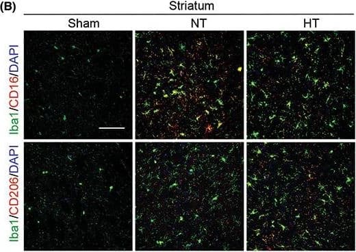
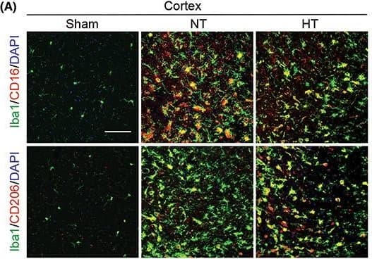
Detection of Mouse MMR/CD206/Mannose Receptor by Immunohistochemistry Hypothermia promotes a shift of microglia/macrophages towards an anti‐inflammatory phenotype in aged female mice 7 days after ischemic stroke. Mice were subjected to distal MCAO (dMCAO) or sham surgery. Normothermia (NT) or hypothermia (HT) was induced for 50 min immediately after dMCAO. Coronal brain sections were stained for Iba‐1 (a microglial/macrophage marker) and CD206 (an anti‐inflammatory marker) or CD16 (a pro‐inflammatory marker). (A) Representative images of Iba1/CD16 and Iba1/CD206 immunofluorescence staining in the ipsilateral peri‐infarct cortex (CTX) regions. (B) Representative images showing Iba1/CD16 and Iba1/CD206 immunofluorescence staining in the ipsilateral peri‐infarct striatum (STR) regions. (C) Representative magnified 3‐dimensional images of Iba1/CD16 and Iba1/CD206 staining. (D) Quantification of the total number of Iba1+ cells (upper panel), Iba1+/CD16+ cells (middle panel), and Iba1+/CD206+ cells (lower panel). The number of double‐positive cells was expressed as the number over 100 Iba1+ cells. n = 5 for sham (S), n = 6 for NT and n = 6 for HT. (E) Pearson correlation analysis of NeuN+ cells with Iba1+/CD206+ cells in the CTX region, n = 6 per group. (F) Pearson correlation analysis of beta ‐APP+ cells with Iba1+/CD206+ cells in the CTX region, n = 6 per group. Scale bar: 50 μm. Data are shown as mean ± SD. *p < 0.05, **p < 0.01 NT vs. HT, ns, no significance. Image collected and cropped by CiteAb from the following open publication (https://pubmed.ncbi.nlm.nih.gov/36341958), licensed under a CC-BY license. Not internally tested by R&D Systems.
 View Larger
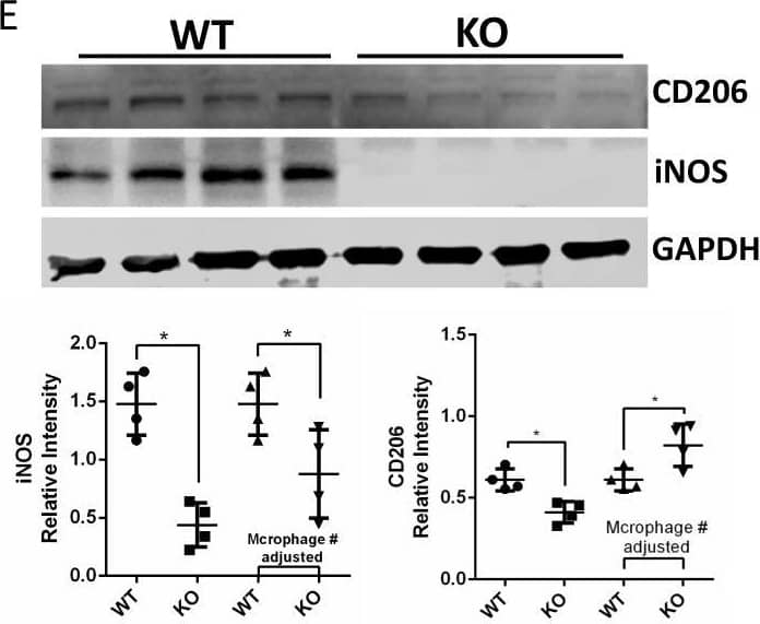
Detection of Mouse MMR/CD206/Mannose Receptor by Western Blot HCK knockout reduced kidney fibrosis in uIRIx model with regulation of macrophage activation.A Schematic diagram of strategy of uIRIx models. B Urine ACR and serum BUN for WT and HCK KO uIRIx mice. C Representative images of H&E, COL1A1 IF and Masson trichrome staining from WT and KO uIRIx kidneys. Morphometric quantification (n = 5 animals; 5 random fields/animal) of the COL1A1 (top) and Masson (lower) positive area. D Representative IF staining images and quantification of from kidneys of HCK and F4/80 macrophage from WT and HCK KO uIRIx kidneys. E Western blot and quantification of M1 and M2 macrophage markers from WT and HCK KO mice kidneys at 28 days post uIRIx. F Representative IF staining images and quantification of LC3 in macrophages from WT & HCK KO uIRIx kidneys. B, C and E*p < 0.05 with t test. Source data are provided as a Source Data file. Image collected and cropped by CiteAb from the following open publication (https://pubmed.ncbi.nlm.nih.gov/37463911), licensed under a CC-BY license. Not internally tested by R&D Systems.
 View Larger
Detection of Mouse MMR/CD206/Mannose Receptor by Immunohistochemistry Hypothermia promotes a shift of microglia/macrophages towards an anti‐inflammatory phenotype in aged female mice 7 days after ischemic stroke. Mice were subjected to distal MCAO (dMCAO) or sham surgery. Normothermia (NT) or hypothermia (HT) was induced for 50 min immediately after dMCAO. Coronal brain sections were stained for Iba‐1 (a microglial/macrophage marker) and CD206 (an anti‐inflammatory marker) or CD16 (a pro‐inflammatory marker). (A) Representative images of Iba1/CD16 and Iba1/CD206 immunofluorescence staining in the ipsilateral peri‐infarct cortex (CTX) regions. (B) Representative images showing Iba1/CD16 and Iba1/CD206 immunofluorescence staining in the ipsilateral peri‐infarct striatum (STR) regions. (C) Representative magnified 3‐dimensional images of Iba1/CD16 and Iba1/CD206 staining. (D) Quantification of the total number of Iba1+ cells (upper panel), Iba1+/CD16+ cells (middle panel), and Iba1+/CD206+ cells (lower panel). The number of double‐positive cells was expressed as the number over 100 Iba1+ cells. n = 5 for sham (S), n = 6 for NT and n = 6 for HT. (E) Pearson correlation analysis of NeuN+ cells with Iba1+/CD206+ cells in the CTX region, n = 6 per group. (F) Pearson correlation analysis of beta ‐APP+ cells with Iba1+/CD206+ cells in the CTX region, n = 6 per group. Scale bar: 50 μm. Data are shown as mean ± SD. *p < 0.05, **p < 0.01 NT vs. HT, ns, no significance. Image collected and cropped by CiteAb from the following open publication (https://pubmed.ncbi.nlm.nih.gov/36341958), licensed under a CC-BY license. Not internally tested by R&D Systems.
Preparation and Storage
- 12 months from date of receipt, -20 to -70 °C as supplied.
- 1 month, 2 to 8 °C under sterile conditions after reconstitution.
- 6 months, -20 to -70 °C under sterile conditions after reconstitution.
Background: MMR/CD206
The mouse Macrophage Mannose Receptor (MMR), also known as CD206 and MRC1 (mannose receptor C, type 1), is a 175 kDa scavenger receptor that is expressed on tissue macrophages, myeloid dendritic cells, and liver and lymphatic endothelial cells (1). It belongs to a family of receptors sharing similar protein structure that also includes DEC205, phospholipase A2 receptor, and Endo180 (2, 3). The mouse MMR protein is synthesized as a 1456 amino acid (aa) precursor that contains a 19 aa signal sequence, a 1369 aa extracellular region, a 21 aa transmembrane segment and a 47 aa cytoplasmic domain (4). Its extracellular region is composed of an N-terminal cysteine-rich domain, followed by a single fibronectin type II repeat, and eight C-type lectin carbohydrate recognition domains (CRD) (3‑5). Mouse to human, the extracellular region is 82% aa identical. The cysteine-rich domain mediates recognition of sulfated N-acetylgalactosamine, which occurs on some extracellular matrix proteins and is the terminal sugar of the unusual oligosaccharides present on pituitary hormones such as lutropin and thyrotropin (6). Several of the CRDs participate in the Ca2+-dependent recognition of carbohydrates showing a preference for branched sugars with terminal mannose, fucose or N‑acetylglucosamine (7). The cytoplasmic domain of MMR includes a tyrosine-based motif for internalization in clathrin-coated vesicles. Once internalized, ligands are released following acidification of phagosomes or endosomes, and the receptor recycles to the cell surface (3, 8). MMR mediates phagocytosis upon binding to target structures that occur on a variety of pathogenic microorganisms including Gram-negative and Gram-positive bacteria, yeasts, parasites, and mycobacteria. MMR also functions to maintain homeostasis through the endocytosis of potentially harmful glycoproteins associated with inflammation (2, 3).
- East, L. and C. Isake (2002) Biochim. Biophys. Acta 1572:364.
- Chieppa, M. et al. (2003) J. Immunol. 171:4552.
- Figdor, C. et al. (2002) Nat. Rev. Immunol. 2:77.
- Harris, N. et al. (1992) Blood 80:2363.
- Taylor, M. et al. (1990) J. Biol. Chem. 265:12156.
- Leteux, C. et al. (2000) J. Exp. Med. 191:1117.
- Martinez-Pomares, L. et al. (2001) Immunobiology 204:527.
- Feinberg, H. et al. (2000) J. Biol. Chem. 275:21539.
Product Datasheets
Citations for Mouse MMR/CD206 Antibody
R&D Systems personnel manually curate a database that contains references using R&D Systems products. The data collected includes not only links to publications in PubMed, but also provides information about sample types, species, and experimental conditions.
194
Citations: Showing 1 - 10
Filter your results:
Filter by:
-
Osteopontin Augments M2 Microglia Response and Separates M1- and M2-Polarized Microglial Activation in Permanent Focal Cerebral Ischemia
Authors: A Ladwig, HL Walter, J Hucklenbro, A Willuweit, KJ Langen, GR Fink, MA Rueger, M Schroeter
Mediators Inflamm., 2017-09-20;2017(0):7189421.
-
A Distinct Microglial Cell Population Expressing Both CD86 and CD206 Constitutes a Dominant Type and Executes Phagocytosis in Two Mouse Models of Retinal Degeneration
Authors: Zhang, Y;Park, YS;Kim, IB;
International journal of molecular sciences
-
Eluted 25-hydroxyvitamin D3 from radially aligned nanofiber scaffolds enhances cathelicidin production while reducing inflammatory response in human immune system-engrafted mice
Authors: Chen S, Ge L, Wang H et al.
Acta Biomater
-
Epigenetic regulation of brain region-specific microglia clearance activity
Authors: P Ayata, A Badimon, HJ Strasburge, MK Duff, SE Montgomery, YE Loh, A Ebert, AA Pimenova, BR Ramirez, AT Chan, JM Sullivan, I Purushotha, JR Scarpa, AM Goate, M Busslinger, L Shen, B Losic, A Schaefer
Nat. Neurosci., 2018-07-23;21(8):1049-1060.
-
Docosahexaenoic acid decreased neuroinflammation in rat pups after controlled cortical impact
Authors: Schober ME, Requena DF, Casper TC Et al.
Exp Neurol
-
Compression Decreases Anatomical and Functional Recovery and Alters Inflammation after Contusive Spinal Cord Injury
Authors: MB Orr, J Simkin, WM Bailey, NH Kadambi, AL McVicar, AK Veldhorst, J Gensel
J. Neurotrauma, 2017-06-14;0(0):.
-
Symbiotic Macrophage-Glioma Cell Interactions Reveal Synthetic Lethality in PTEN-Null Glioma
Authors: Chen P, Zhao D, Li J et al.
Cancer Cell
-
MiR-101a loaded extracellular nanovesicles as bioactive carriers for cardiac repair
Authors: Wang J, Lee CJ, Deci MB et al.
Nanomedicine
-
Collectin-11 promotes cancer cell proliferation and tumor growth
Authors: JX Wang, B Cao, N Ma, KY Wu, WB Chen, W Wu, X Dong, CF Liu, YF Gao, TY Diao, XY Min, Q Yong, ZF Li, W Zhou, K Li
JCI Insight, 2023-03-08;8(5):.
-
Unraveling the transcriptional determinants of Liver Sinusoidal Endothelial Cell specialization
Authors: de Haan W, Oie CI, Benkheil M et al.
Am. J. Physiol. Gastrointest. Liver Physiol.
-
Mechanistic Studies of Gypenosides in Microglial State Transition and its Implications in Depression-Like Behaviors: Role of TLR4/MyD88/NF-kappa B Signaling
Authors: Li-Hua Cao, Yuan-Yuan Zhao, Ming Bai, David Geliebter, Jan Geliebter, Raj Tiwari et al.
Frontiers in Pharmacology
-
Augmentation of a neuroprotective myeloid state by hematopoietic cell transplantation
Authors: MM Mader, A Napole, D Wu, Y Shibuya, A Scavetti, A Foltz, M Atkins, O Hahn, Y Yoo, R Danziger, C Tan, T Wyss-Coray, L Steinman, M Wernig
bioRxiv : the preprint server for biology, 2023-03-12;0(0):.
-
A maresin 1/ROR alpha/12-lipoxygenase autoregulatory circuit prevents inflammation and progression of nonalcoholic steatohepatitis
Authors: Han YH, Shin KO, Kim JY et al.
J. Clin. Invest.
-
STING orchestrates microglia polarization via interaction with LC3 in autophagy after ischemia
Authors: Kong, L;Xu, P;Shen, N;Li, W;Li, R;Tao, C;Wang, G;Zhang, Y;Sun, W;Hu, W;Liu, X;
Cell death & disease
Species: Mouse
Sample Types: Cell Lysates, Tissue Homogenates, Whole Tissue
Applications: Immunohistochemistry, Western Blot -
Dermal macrophages control tactile perception under physiological conditions via NGF signaling
Authors: Tanaka, T;Isonishi, A;Banja, M;Yamamoto, R;Sonobe, M;Okuda-Ashitaka, E;Furue, H;Okuda, H;Tatsumi, K;Wanaka, A;
Scientific reports
Species: Transgenic Mouse
Sample Types: Tissue Homogenates
Applications: Western Blot -
Matrix-bound nanovesicles alleviate particulate-induced periprosthetic osteolysis
Authors: Liao, R;Dewey, MJ;Rong, J;Johnson, SA;D'Angelo, WA;Hussey, GS;Badylak, SF;
Science advances
Species: Mouse
Sample Types: Whole Tissue
Applications: Immunohistochemistry -
Inhibiting Ca2+ channels in Alzheimer's disease model mice relaxes pericytes, improves cerebral blood flow and reduces immune cell stalling and hypoxia
Authors: Korte, N;Barkaway, A;Wells, J;Freitas, F;Sethi, H;Andrews, SP;Skidmore, J;Stevens, B;Attwell, D;
Nature neuroscience
Species: Mouse
Sample Types: Whole Tissue
Applications: Immunohistochemistry -
Inhalable SPRAY nanoparticles by modular peptide assemblies reverse alveolar inflammation in lethal Gram-negative bacteria infection
Authors: Chen, D;Zhou, Z;Kong, N;Xu, T;Liang, J;Xu, P;Yao, B;Zhang, Y;Sun, Y;Li, Y;Wu, B;Yang, X;Wang, H;
Science advances
Species: Mouse
Sample Types: Whole Cells, Whole Tissue
Applications: Immunohistochemistry, Immunocytochemistry -
Tumor necrosis factor-?-treated human adipose-derived stem cells enhance inherent radiation tolerance and alleviate in vivo radiation-induced capsular contracture
Authors: Sutthiwanjampa, C;Kang, SH;Kim, MK;Hwa Choi, J;Kim, HK;Woo, SH;Bae, TH;Kim, WJ;Kang, SH;Park, H;
Journal of advanced research
Species: Human
Sample Types: Tissue Array
Applications: IHC-Pr -
Adjudin protects blood-brain barrier integrity and attenuates neuroinflammation following intracerebral hemorrhage in mice
Authors: Su, Q;Su, C;Zhang, Y;Guo, Y;Liu, Y;Liu, Y;Yong, VW;Xue, M;
International immunopharmacology
Species: Mouse
Sample Types: Whole Tissue
Applications: Immunohistochemistry -
Myeloid cell replacement is neuroprotective in chronic experimental autoimmune encephalomyelitis
Authors: Mader, MM;Napole, A;Wu, D;Atkins, M;Scavetti, A;Shibuya, Y;Foltz, A;Hahn, O;Yoo, Y;Danziger, R;Tan, C;Wyss-Coray, T;Steinman, L;Wernig, M;
Nature neuroscience
Species: Mouse
Sample Types: Whole Tissue
Applications: IHC/IF -
Intracerebellar injection of monocytic immature myeloid cells prevents the adverse effects caused by stereotactic surgery in a model of cerebellar neurodegeneration
Authors: Del Pilar, C;Garrido-Matilla, L;Del Pozo-Filíu, L;Lebrón-Galán, R;Arias, RF;Clemente, D;Alonso, JR;Weruaga, E;Díaz, D;
Journal of neuroinflammation
Species: Mouse
Sample Types: Whole Tissue
Applications: IHC -
Signaling events at TMEM doorways provide potential targets for inhibiting breast cancer dissemination
Authors: Surve, CR;Duran, CL;Ye, X;Chen, X;Lin, Y;Harney, AS;Wang, Y;Sharma, VP;Stanley, ER;McAuliffe, JC;Entenberg, D;Oktay, MH;Condeelis, JS;
bioRxiv : the preprint server for biology
Species: Mouse
Sample Types: Whole Tissue
Applications: IHC -
M2a macrophages facilitate resolution of chemically-induced colitis in TLR4-SNP mice
Authors: Vlk, AM;Prantner, D;Shirey, KA;Perkins, DJ;Buzza, MS;Thumbigere-Math, V;Keegan, AD;Vogel, SN;
mBio
Species: Mouse
Sample Types: Cell Culture Supernates
Applications: Western Blot -
Differential Effects of Regulatory T Cells in the Meninges and Spinal Cord of Male and Female Mice with Neuropathic Pain
Authors: Fiore, NT;Keating, BA;Chen, Y;Williams, SI;Moalem-Taylor, G;
Cells
Species: Mouse
Sample Types: Whole Tissue
Applications: IHC -
Manganese-Implanted Titanium Modulates the Crosstalk between Bone Marrow Mesenchymal Stem Cells and Macrophages to Improve Osteogenesis
Authors: Ye, K;Zhang, X;Shangguan, L;Liu, X;Nie, X;Qiao, Y;
Journal of functional biomaterials
Species: Mouse
Sample Types: Whole Cells
Applications: ICC -
Circadian clock regulator Bmal1 gates axon regeneration via Tet3 epigenetics in mouse sensory neurons
Authors: Halawani, D;Wang, Y;Ramakrishnan, A;Estill, M;He, X;Shen, L;Friedel, RH;Zou, H;
Nature communications
Species: Transgenic Mouse
Sample Types: Whole Tissue
Applications: Immunohistochemistry -
The Single-Dose Application of Interleukin-4 Ameliorates Secondary Brain Damage in the Early Phase after Moderate Experimental Traumatic Brain Injury in Mice
Authors: Walter, J;Mende, J;Hutagalung, S;Alhalabi, OT;Grutza, M;Zheng, G;Skutella, T;Unterberg, A;Zweckberger, K;Younsi, A;
International journal of molecular sciences
Species: Mouse
Sample Types: Whole Tissue
Applications: IHC -
Fetal Muse-based therapy prevents lethal radio-induced gastrointestinal syndrome by intestinal regeneration
Authors: Honorine Dushime, Stéphanie G. Moreno, Christine Linard, Annie Adrait, Yohann Couté, Juliette Peltzer et al.
Stem Cell Research & Therapy
-
MicroRNA-124-3p Attenuated Retinal Neovascularization in Oxygen-Induced Retinopathy Mice by Inhibiting the Dysfunction of Retinal Neuroglial Cells through STAT3 Pathway
Authors: Hong Y, Wang Y, Cui Y et al.
International journal of molecular sciences
-
Macrophage fusion event as one prerequisite for inorganic nanoparticle-induced antitumor response
Authors: Chen S, Xing Z, Geng M et al.
Science advances
-
HCK induces macrophage activation to promote renal inflammation and fibrosis via suppression of autophagy
Authors: Chen M, Menon MC, Wang W et al.
Nature communications
-
An invasive zone in human liver cancer identified by Stereo-seq promotes hepatocyte-tumor cell crosstalk, local immunosuppression and tumor progression
Authors: Wu, L;Yan, J;Bai, Y;Chen, F;Zou, X;Xu, J;Huang, A;Hou, L;Zhong, Y;Jing, Z;Yu, Q;Zhou, X;Jiang, Z;Wang, C;Cheng, M;Ji, Y;Hou, Y;Luo, R;Li, Q;Wu, L;Cheng, J;Wang, P;Guo, D;Huang, W;Lei, J;Liu, S;Yan, Y;Chen, Y;Liao, S;Li, Y;Sun, H;Yao, N;Zhang, X;Zhang, S;Chen, X;Yu, Y;Li, Y;Liu, F;Wang, Z;Zhou, S;Yang, H;Yang, S;Xu, X;Liu, L;Gao, Q;Tang, Z;Wang, X;Wang, J;Fan, J;Liu, S;Yang, X;Chen, A;Zhou, J;
Cell research
Species: Human
Sample Types: Whole Tissue
Applications: IHC -
Oleoylethanolamide Treatment Modulates Both Neuroinflammation and Microgliosis, and Prevents Massive Leukocyte Infiltration to the Cerebellum in a Mouse Model of Neuronal Degeneration
Authors: Pérez-Martín, E;Pérez-Revuelta, L;Barahona-López, C;Pérez-Boyero, D;Alonso, JR;Díaz, D;Weruaga, E;
International journal of molecular sciences
Species: Mouse
Sample Types: Whole Tissue
Applications: IHC -
CD301b+ macrophage: the new booster for activating bone regeneration in periodontitis treatment
Authors: Can Wang, Qin Zhao, Chen Chen, Jiaojiao Li, Jing Zhang, Shuyuan Qu et al.
International Journal of Oral Science
-
Age-related neuroimmune signatures in dorsal root ganglia of a Fabry disease mouse model
Authors: Choconta JL, Labi V, Dumbraveanu C et al.
Immunity & ageing : I & A
-
iPSC-sEVs alleviate microglia senescence to protect against ischemic stroke in aged mice
Authors: Xinyu Niu, Yuguo Xia, Lei Luo, Yu Chen, Ji Yuan, Juntao Zhang et al.
Materials Today Bio
-
Combining three independent pathological stressors induces a heart failure with preserved ejection fraction phenotype
Authors: Li Y, Kubo H, Yu D et al.
American journal of physiology. Heart and circulatory physiology
-
Meningeal origins and dynamics of perivascular fibroblast development on the mouse cerebral vasculature
Authors: HE Jones, V Coelho-San, SK Bonney, KA Abrams, AY Shih, JA Siegenthal
bioRxiv : the preprint server for biology, 2023-03-23;0(0):.
Species: Mouse
Sample Types: Whole Tissue
Applications: IHC -
Circadian regulator CLOCK promotes tumor angiogenesis in glioblastoma
Authors: L Pang, M Dunterman, W Xuan, A Gonzalez, Y Lin, WH Hsu, F Khan, RS Hagan, WA Muller, AB Heimberger, P Chen
Cell Reports, 2023-02-14;42(2):112127.
Species: Mouse
Sample Types: Whole Tissue
Applications: IHC -
Regulating macrophage-MSC interaction to optimize BMP-2-induced osteogenesis in the local microenvironment
Authors: F Jiang, X Qi, X Wu, S Lin, J Shi, W Zhang, X Jiang
Bioactive materials, 2023-02-11;25(0):307-318.
Species: Mouse
Sample Types: Whole Cells
Applications: ICC -
Implantation and tracing of green fluorescent protein-expressing adipose-derived stem cells in peri-implant capsular fibrosis
Authors: Bo-Yoon Park, Dirong Wu, Kyoo-Ri Kwon, Mi-Jin Kim, Tae-Gon Kim, Jun-Ho Lee et al.
Stem Cell Research & Therapy
-
CD206+ macrophages transventricularly infiltrate the early embryonic cerebral wall to differentiate into microglia
Authors: Y Hattori, D Kato, F Murayama, S Koike, H Asai, A Yamasaki, Y Naito, A Kawaguchi, H Konishi, M Prinz, T Masuda, H Wake, T Miyata
Cell Reports, 2023-02-07;42(2):112092.
Species: Mouse
Sample Types: Whole Tissue
Applications: IHC -
White-light crosslinkable milk protein bioadhesive with ultrafast gelation for first-aid wound treatment
Authors: Q Zhu, X Zhou, Y Zhang, D Ye, K Yu, W Cao, L Zhang, H Zheng, Z Sun, C Guo, X Hong, Y Zhu, Y Zhang, Y Xiao, TG Valencak, T Ren, D Ren
Biomaterials research, 2023-02-03;27(1):6.
Species: Mouse
Sample Types: Whole Tissue
Applications: IHC -
Dermal macrophages set pain sensitivity by modulating the amount of tissue NGF through an SNX25-Nrf2 pathway
Authors: T Tanaka, H Okuda, A Isonishi, Y Terada, M Kitabatake, T Shinjo, K Nishimura, S Takemura, H Furue, T Ito, K Tatsumi, A Wanaka
Nature Immunology, 2023-01-26;0(0):.
Species: Mouse
Sample Types: Tissue Homogenates
Applications: Western Blot -
Protective effects of blocking PD-1 pathway on retinal ganglion cells in a mouse model of chronic ocular hypertension
Authors: Siqi Sheng, Yixian Ma, Yue Zou, Fangyuan Hu, Ling Chen
Frontiers in Immunology
-
Selective brain hypothermia attenuates focal cerebral ischemic injury and improves long‐term neurological outcome in aged female mice
Authors: Liqiang Liu, Jia Liu, Ming Li, Junxuan Lyu, Wei Su, Shejun Feng et al.
CNS Neuroscience & Therapeutics
-
Inhibiting phosphatase and actin regulator 1 expression is neuroprotective in the context of traumatic brain injury
Authors: Heng-Li Tian, Zhi-Ming Zhang, Shi-Wen Ding, Yao Jing, Shi-Ming Zhang, Shi-Wen Chen et al.
Neural Regeneration Research
-
Antibody-Mediated Delivery of VEGF-C Promotes Long-Lasting Lymphatic Expansion That Reduces Recurrent Inflammation
Authors: N Cousin, S Bartel, J Scholl, C Tacconi, A Egger, G Thorhallsd, D Neri, LC Dieterich, M Detmar
Cells, 2022-12-31;12(1):.
Species: Mouse
Sample Types: Whole Tissue
Applications: IHC -
An SPM-Enriched Marine Oil Supplement Shifted Microglia Polarization toward M2, Ameliorating Retinal Degeneration in rd10 Mice
Authors: Lorena Olivares-González, Sheyla Velasco, Idoia Gallego, Marina Esteban-Medina, Gustavo Puras, Carlos Loucera et al.
Antioxidants (Basel)
-
Infiltration of meningeal macrophages into the Virchow–Robin space after ischemic stroke in rats: Correlation with activated PDGFR-beta -positive adventitial fibroblasts
Authors: Tae-Ryong Riew, Ji-Won Hwang, Xuyan Jin, Hong Lim Kim, Mun-Yong Lee
Frontiers in Molecular Neuroscience
-
Long-term functional regeneration of radiation-damaged salivary glands through delivery of a neurogenic hydrogel
Authors: J Li, S Sudiwala, L Berthoin, S Mohabbat, EA Gaylord, H Sinada, N Cruz Pache, JC Chang, O Jeon, IMA Lombaert, AJ May, E Alsberg, CS Bahney, SM Knox
Science Advances, 2022-12-21;8(51):eadc8753.
Species: Mouse
Sample Types: Whole Tissue
Applications: IHC/IF -
Tumor necrosis factor-alpha -primed mesenchymal stem cell-derived exosomes promote M2 macrophage polarization via Galectin-1 and modify intrauterine adhesion on a novel murine model
Authors: Jingman Li, Yuchen Pan, Jingjing Yang, Jiali Wang, Qi Jiang, Huan Dou et al.
Frontiers in Immunology
-
Interleukin-4 promotes microglial polarization toward a neuroprotective phenotype after retinal ischemia/reperfusion injury
Authors: D Chen, C Peng, XM Ding, Y Wu, CJ Zeng, L Xu, WY Guo
Oncogene, 2022-12-01;17(12):2755-2760.
Species: Mouse
Sample Types: Whole Tissue
Applications: IHC -
Targeting the bicarbonate transporter SLC4A4 overcomes immunosuppression and immunotherapy resistance in pancreatic cancer
Authors: Cappellesso F, Orban MP, Shirgaonkar N et al.
Nature cancer
-
IL-33/ST2 axis promotes remodeling of the extracellular matrix and drives protective microglial responses in the mouse model of perioperative neurocognitive disorders
Authors: S Li, H Liu, Y Qian, L Jiang, S Liu, Y Liu, C Liu, X Gu
International immunopharmacology, 2022-11-26;114(0):109479.
Species: Mouse
Sample Types: Tissue Homogenates
Applications: Western Blot -
Constitutively active microglial populations limit anorexia induced by the food contaminant deoxynivalenol
Authors: S Gaige, R Barbouche, M Barbot, S Boularand, M Dallaporta, A Abysique, JD Troadec
Journal of Neuroinflammation, 2022-11-19;19(1):280.
Species: Mouse
Sample Types: Whole Tissue
Applications: IHC -
Reversible Myc hypomorphism identifies a key Myc-dependency in early cancer evolution
Authors: NM Sodir, L Pellegrine, RM Kortlever, T Campos, YW Kwon, S Kim, D Garcia, A Perfetto, P Anastasiou, LB Swigart, MJ Arends, TD Littlewood, GI Evan
Nature Communications, 2022-11-09;13(1):6782.
Species: Mouse
Sample Types: Whole Tissue
Applications: IHC -
HRas and Myc synergistically induce cell cycle progression and apoptosis of murine cardiomyocytes
Authors: Aleksandra Boikova, Megan J. Bywater, Gregory A. Quaife-Ryan, Jasmin Straube, Lucy Thompson, Camilla Ascanelli et al.
Frontiers in Cardiovascular Medicine
-
Multiplex immunohistochemistry reveals cochlear macrophage heterogeneity and local auditory nerve inflammation in cisplatin-induced hearing loss
Authors: Mai Mohamed Bedeir, Yuzuru Ninoyu, Takashi Nakamura, Takahiro Tsujikawa, Shigeru Hirano
Frontiers in Neurology
-
Fibrocytes boost tumor-supportive phenotypic switches in the lung cancer niche via the endothelin system
Authors: A Weigert, X Zheng, A Nenzel, K Turkowski, S Günther, E Strack, E Sirait-Fis, E Elwakeel, IM Kur, VS Nikam, C Valasaraja, H Winter, A Wissgott, R Voswinkel, F Grimminger, B Brüne, W Seeger, SS Pullamsett, R Savai
Nature Communications, 2022-10-14;13(1):6078.
Species: Mouse
Sample Types: Whole Tissue
Applications: IHC -
Cancer cell autophagy, reprogrammed macrophages, and remodeled vasculature in glioblastoma triggers tumor immunity
Authors: Agnieszka Chryplewicz, Julie Scotton, Mélanie Tichet, Anoek Zomer, Ksenya Shchors, Johanna A. Joyce et al.
Cancer Cell
-
Ginsenoside Rg3-enriched Korean red ginseng extract attenuates Non-Alcoholic Fatty Liver Disease by way of suppressed VCAM-1 expression in liver sinusoidal endothelium
Authors: Lee S, Baek S, Lee Y et al.
Journal of Ginseng Research
Species: Mouse
Sample Types: Whole Tissue
Applications: Immunohistochemistry -
Chuanzhitongluo regulates microglia polarization and inflammatory response in acute ischemic stroke
Authors: Q Wang, B Han, X Man, H Gu, J Sun
Brain research bulletin, 2022-09-21;190(0):97-104.
Species: Mouse
Sample Types: Whole Tissue
Applications: IHC -
Large extracellular vesicles secreted by human iPSC-derived MSCs ameliorate tendinopathy via regulating macrophage heterogeneity
Authors: T Ye, Z Chen, J Zhang, L Luo, R Gao, L Gong, Y Du, Z Xie, B Zhao, Q Li, Y Wang
Oncogene, 2022-08-26;21(0):194-208.
Species: Mouse
Sample Types: Whole Cells
Applications: ICC -
Transforming growth factor-beta1 protects against LPC-induced cognitive deficit by attenuating pyroptosis of microglia via NF-kappaB/ERK1/2 pathways
Authors: Y Xie, X Chen, Y Li, S Chen, S Liu, Z Yu, W Wang
Journal of Neuroinflammation, 2022-07-28;19(1):194.
Species: Mouse
Sample Types: Cell Lysates
Applications: Western Blot -
Rationally designed bioactive milk-derived protein scaffolds enhanced new bone formation
Authors: MS Lee, J Jeon, S Park, J Lim, HS Yang
Oncogene, 2022-06-16;20(0):368-380.
Species: Mouse
Sample Types: Whole Tissue
Applications: IHC -
FGF-2 signaling in nasopharyngeal carcinoma modulates pericyte-macrophage crosstalk and metastasis
Authors: Y Wang, Q Sun, Y Ye, X Sun, S Xie, Y Zhan, J Song, X Fan, B Zhang, M Yang, L Lv, K Hosaka, Y Yang, G Nie
JCI Insight, 2022-05-23;0(0):.
Species: Mouse
Sample Types: Whole Tissue
Applications: IHC -
Interleukin 13 promotes long-term recovery after ischemic stroke by inhibiting the activation of STAT3
Authors: D Chen, J Li, Y Huang, P Wei, W Miao, Y Yang, Y Gao
Journal of Neuroinflammation, 2022-05-16;19(1):112.
Species: Mouse
Sample Types: Whole Tissue
Applications: IHC -
Repetitive transcranial magnetic stimulation exerts anti-inflammatory effects via modulating glial activation in mice with chronic unpredictable mild stress-induced depression
Authors: C Zuo, H Cao, F Feng, G Li, Y Huang, L Zhu, Z Gu, Y Yang, J Chen, Y Jiang, F Wang
International immunopharmacology, 2022-04-30;109(0):108788.
Species: Mouse
Sample Types: Tissue Homogenates, Whole Tissue
Applications: IF, Western Blot -
Specification of CNS macrophage subsets occurs postnatally in defined niches
Authors: T Masuda, L Amann, G Monaco, R Sankowski, O Staszewski, M Krueger, F Del Gaudio, L He, N Paterson, E Nent, F Fernández-, A Yamasaki, M Frosch, M Fliegauf, LFP Bosch, H Ulupinar, N Hagemeyer, D Schreiner, C Dorrier, M Tsuda, C Grothe, A Joutel, R Daneman, C Betsholtz, U Lendahl, KP Knobeloch, T Lämmermann, J Priller, K Kierdorf, M Prinz
Nature, 2022-04-20;604(7907):740-748.
Species: Mouse, Transgenic Mouse
Sample Types: Whole Tissue
Applications: IHC -
Oncogenic Vav1-Myo1f induces therapeutically targetable macrophage-rich tumor microenvironment in peripheral T�cell lymphoma
Authors: JR Cortes, I Filip, R Albero, JA Patiño-Gal, SA Quinn, WW Lin, AP Laurent, BB Shih, JA Brown, AJ Cooke, A Mackey, J Einson, S Zairis, A Rivas-Delg, MA Laginestra, S Pileri, E Campo, G Bhagat, AA Ferrando, R Rabadan, T Palomero
Cell Reports, 2022-04-19;39(3):110695.
Species: Mouse
Sample Types: Whole Cells, Whole Tissue
Applications: Flow Cytometry, IHC -
Specification of fetal liver endothelial progenitors to functional zonated adult sinusoids requires c-Maf induction
Authors: Jesus Maria Gómez-Salinero, Franco Izzo, Yang Lin, Sean Houghton, Tomer Itkin, Fuqiang Geng et al.
Cell Stem Cell
-
Targeted Accumulation of Macrophages Induced by Microbeam Irradiation in a Tissue-Dependent Manner
Authors: V Trappetti, J Fazzari, C Fernandez-, L Smyth, M Potez, N Shintani, B de Breuyn, OA Martin, V Djonov
Biomedicines, 2022-03-22;10(4):.
Species: Mouse
Sample Types: Whole Tissue
Applications: IHC -
A Novel Staining Method for Detection of Brain Perivascular Injuries Induced by Nanoparticle: Periodic Acid-Schiff and Immunohistochemical Double-Staining
Authors: Atsuto Onoda, Shin Hagiwara, Natsuko Kubota, Shinya Yanagita, Ken Takeda, Masakazu Umezawa
Frontiers in Toxicology
-
A bioactive gypenoside (GP-14) alleviates neuroinflammation and blood brain barrier (BBB) disruption by inhibiting the NF-kappaB signaling pathway in a mouse high-altitude cerebral edema (HACE) model
Authors: Y Geng, J Yang, X Cheng, Y Han, F Yan, C Wang, X Jiang, X Meng, M Fan, M Zhao, L Zhu
International immunopharmacology, 2022-03-14;107(0):108675.
Species: Mouse
Sample Types: Whole Tissue
Applications: IHC -
MYC Levels Regulate Metastatic Heterogeneity in Pancreatic Adenocarcinoma
Authors: Ravikanth Maddipati, Robert J. Norgard, Timour Baslan, Komal S. Rathi, Amy Zhang, Asal Saeid et al.
Cancer Discovery
-
A muscle cell‐macrophage axis involving matrix metalloproteinase 14 facilitates extracellular matrix remodeling with mechanical loading
Authors: Bailey D. Peck, Kevin A. Murach, R. Grace Walton, Alexander J. Simmons, Douglas E. Long, Kate Kosmac et al.
The FASEB Journal
-
miR-204-containing exosomes ameliorate GVHD-associated dry eye disease
Authors: T Zhou, C He, P Lai, Z Yang, Y Liu, H Xu, X Lin, B Ni, R Ju, W Yi, L Liang, D Pei, CE Egwuagu, X Liu
Science Advances, 2022-01-12;8(2):eabj9617.
Species: Mouse
Sample Types: Whole Tissue
Applications: IHC -
An HDAC6 inhibitor reverses chemotherapy-induced mechanical hypersensitivity via an IL-10 and macrophage dependent pathway
Authors: J Zhang, J Ma, RT Trinh, CJ Heijnen, A Kavelaars
Brain, Behavior, and Immunity, 2021-12-13;100(0):287-296.
Species: Mouse
Sample Types: Whole Tissue
Applications: IHC -
Characterization of macrophage infiltration and polarization by double fluorescence immunostaining in mouse colonic mucosa
Authors: María López Chiloeches, Anna Bergonzini, Teresa Frisan, Océane C.B. Martin
STAR Protocols
-
Mad2 Induced Aneuploidy Contributes to Eml4-Alk Driven Lung Cancer by Generating an Immunosuppressive Environment
Authors: Kristina Alikhanyan, Yuanyuan Chen, Kalman Somogyi, Simone Kraut, Rocio Sotillo
Cancers (Basel)
-
Hypothermia modulates myeloid cell polarization in neonatal hypoxic-ischemic brain injury
Authors: M Seitz, C Köster, M Dzietko, H Sabir, M Serdar, U Felderhoff, I Bendix, J Herz
Journal of Neuroinflammation, 2021-11-13;18(1):266.
Species: Mouse
Sample Types: Whole Tissue
Applications: IHC -
Remote Limb Ischemic Postconditioning Protects Against Ischemic Stroke by Promoting Regulatory T Cells Thriving
Authors: HH Yu, XT Ma, X Ma, M Chen, YH Chu, LJ Wu, W Wang, C Qin, DS Tian
Journal of the American Heart Association, 2021-11-02;10(22):e023077.
Species: Mouse
Sample Types: Whole Cells, Whole Tissue
Applications: ICC, IHC -
Raloxifene Modulates Microglia and Rescues Visual Deficits and Pathology After Impact Traumatic Brain Injury
Authors: Marcia G. Honig, Nobel A. Del Mar, Desmond L. Henderson, Dylan O’Neal, John B. Doty, Rachel Cox et al.
Frontiers in Neuroscience
-
Tissue-resident M2 macrophages directly contact primary sensory neurons in the sensory ganglia after nerve injury
Authors: H Iwai, K Ataka, H Suzuki, A Dhar, E Kuramoto, A Yamanaka, T Goto
Journal of Neuroinflammation, 2021-10-13;18(1):227.
Species: Mouse
Sample Types: Whole Tissue
Applications: IF -
Activating a collaborative innate-adaptive immune response to control metastasis
Authors: Lijuan Sun, Tim Kees, Ana Santos Almeida, Bodu Liu, Xue-Yan He, David Ng et al.
Cancer Cell
-
Regulatory T cells protect against brain damage by alleviating inflammatory response in neuromyelitis optica spectrum disorder
Authors: X Ma, C Qin, M Chen, HH Yu, YH Chu, TJ Chen, DB Bosco, LJ Wu, BT Bu, W Wang, DS Tian
Journal of Neuroinflammation, 2021-09-15;18(1):201.
Species: Mouse
Sample Types: Tissue Homogenates, Whole Tissue
Applications: IHC, Western Blot -
InVision: An optimized tissue clearing approach for three-dimensional imaging and analysis of intact rodent eyes
Authors: Akshay Gurdita, Philip E.B. Nickerson, Neno T. Pokrajac, Arturo Ortín-Martínez, En Leh Samuel Tsai, Lacrimioara Comanita et al.
iScience
-
Neuronal chemokine-like-factor 1 (CKLF1) up-regulation promotes M1 polarization of microglia in rat brain after stroke
Authors: X Zhou, YN Zhang, FF Li, Z Zhang, LY Cui, HY He, X Yan, WB He, HS Sun, ZP Feng, SF Chu, NH Chen
Acta pharmacologica Sinica, 2021-08-12;0(0):.
Species: Rat
Sample Types: Whole Tissue
Applications: IHC -
A transepithelial pathway delivers succinate to macrophages, thus perpetuating their pro-inflammatory metabolic state
Authors: M Fremder, SW Kim, A Khamaysi, L Shimshilas, H Eini-Rider, IS Park, U Hadad, JH Cheon, E Ohana
Cell Reports, 2021-08-10;36(6):109521.
Species: Mouse
Sample Types: Cell Lysates
Applications: Western Blot -
Axonal Injuries Cast Long Shadows: Long Term Glial Activation in Injured and Contralateral Retinas after Unilateral Axotomy
Authors: María José González-Riquelme, Caridad Galindo-Romero, Fernando Lucas-Ruiz, Marina Martínez-Carmona, Kristy T. Rodríguez-Ramírez, José María Cabrera-Maqueda et al.
International Journal of Molecular Sciences
-
Beneficial effects of dietary supplementation with green tea catechins and cocoa flavanols on aging-related regressive changes in the mouse neuromuscular system
Authors: Sílvia Gras, Alba Blasco, Guillem Mòdol-Caballero, Olga Tarabal, Anna Casanovas, Lídia Piedrafita et al.
Aging (Albany NY)
-
Resveratrol Ameliorates Cardiac Remodeling in a Murine Model of Heart Failure With Preserved Ejection Fraction
Authors: L Zhang, J Chen, L Yan, Q He, H Xie, M Chen
Frontiers in Pharmacology, 2021-06-10;12(0):646240.
Species: Mouse
Sample Types: Whole Tissue
Applications: IHC -
Annexin A1 protects against cerebral ischemia-reperfusion injury by modulating microglia/macrophage polarization via FPR2/ALX-dependent AMPK-mTOR pathway
Authors: X Xu, W Gao, L Li, J Hao, B Yang, T Wang, L Li, X Bai, F Li, H Ren, M Zhang, L Zhang, J Wang, D Wang, J Zhang, L Jiao
Journal of Neuroinflammation, 2021-05-22;18(1):119.
Species: Mouse
Sample Types: Whole Cells, Whole Tissue
Applications: ICC, IHC -
Quercetin Attenuates Trauma-Induced Heterotopic Ossification by Tuning Immune Cell Infiltration and Related Inflammatory Insult
Authors: Juehong Li, Ziyang Sun, Gang Luo, Shuo Wang, Haomin Cui, Zhixiao Yao et al.
Frontiers in Immunology
-
Hypoxia-induced miR-210 modulates the inflammatory response and fibrosis upon acute ischemia
Authors: Z Germana, G Simona, L Marialucia, M Biagina, V Christine, F Paola, C Matteo, C Pasquale, M Davide, T Mario, M Massimilia, G Carlo, S Gaia, M Fabio
Cell Death & Disease, 2021-05-01;12(5):435.
Species: Mouse
Sample Types: Whole Tissue
Applications: IHC -
Tanshinone IIA Protects Against Cerebral Ischemia Reperfusion Injury by Regulating Microglial Activation and Polarization via NF-kappa B Pathway
Authors: Zhibing Song, Jingjing Feng, Qian Zhang, Shanshan Deng, Dahai Yu, Yuefan Zhang et al.
Frontiers in Pharmacology
-
High-salt diet downregulates TREM2 expression and blunts efferocytosis of macrophages after acute ischemic stroke
Authors: M Hu, Y Lin, X Men, S Wang, X Sun, Q Zhu, D Lu, S Liu, B Zhang, W Cai, Z Lu
Journal of Neuroinflammation, 2021-04-12;18(1):90.
Species: Mouse
Sample Types: Whole Tissue
Applications: IHC -
Imaging of Inflammation in Spinal Cord Injury: Novel Insights on the Usage of PFC-Based Contrast Agents
Authors: F Garello, M Boido, M Miglietti, V Bitonto, M Zenzola, M Filippi, F Arena, L Consolino, M Ghibaudi, E Terreno
Biomedicines, 2021-04-03;9(4):.
Species: Mouse
Sample Types: Whole Cells, Whole Tissue
Applications: ICC, IHC -
Mutant p53 suppresses innate immune signaling to promote tumorigenesis
Authors: Monisankar Ghosh, Suchandrima Saha, Julie Bettke, Rachana Nagar, Alejandro Parrales, Tomoo Iwakuma et al.
Cancer Cell
-
Minocycline promotes functional recovery in ischemic stroke by modulating microglia polarization through STAT1/STAT6 pathways
Authors: Yunnan Lu, Mingming Zhou, Yun Li, Yan Li, Ye Hua, Yi Fan
Biochemical Pharmacology
-
Epithelial membrane protein 2 (Emp2) modulates innate immune cell population recruitment at the maternal-fetal interface
Authors: A Chu, SY Kok, J Tsui, MC Lin, B Aguirre, M Wadehra
Journal of reproductive immunology, 2021-03-09;145(0):103309.
Species: Mouse
Sample Types: Whole Tissue
Applications: IHC -
Differentiated glioblastoma cells accelerate tumor progression by shaping the tumor microenvironment via CCN1-mediated macrophage infiltration
Authors: A Uneda, K Kurozumi, A Fujimura, K Fujii, J Ishida, Y Shimazu, Y Otani, Y Tomita, Y Hattori, Y Matsumoto, N Tsuboi, K Makino, S Hirano, A Kamiya, I Date
Acta neuropathologica communications, 2021-02-22;9(1):29.
Species: Mouse
Sample Types: Whole Tissue
Applications: IHC -
Bone marrow mesenchymal stem cell-derived exosomal microRNA-125a promotes M2 macrophage polarization in spinal cord injury by downregulating IRF5
Authors: Q Chang, Y Hao, Y Wang, Y Zhou, H Zhuo, G Zhao
Brain research bulletin, 2021-02-17;170(0):199-210.
Species: Mouse
Sample Types: Whole Cells
Applications: Flow Cytometry -
GSK2593074A blocks progression of existing abdominal aortic dilation
Authors: Mitri K. Khoury, Ting Zhou, Huan Yang, Samantha R. Prince, Kartik Gupta, Amelia R. Stranz et al.
JVS-Vascular Science
-
Astragaloside IV promotes microglia/macrophages M2 polarization and enhances neurogenesis and angiogenesis through PPAR&gamma pathway after cerebral ischemia/reperfusion injury in rats
Authors: L Li, H Gan, H Jin, Y Fang, Y Yang, J Zhang, X Hu, L Chu
International immunopharmacology, 2021-01-08;92(0):107335.
Species: Rat
Sample Types: Whole Tissue
Applications: IHC -
The novel Nrf2 activator CDDO‐EA attenuates cerebral ischemic injury by promoting microglia/macrophage polarization toward M2 phenotype in mice
Authors: Xia Lei, Hanxia Li, Min Li, Qiwei Dong, Huayang Zhao, Zongyong Zhang et al.
CNS Neuroscience & Therapeutics
-
M2 microglial small extracellular vesicles reduce glial scar formation via the miR-124/STAT3 pathway after ischemic stroke in mice
Authors: Z Li, Y Song, T He, R Wen, Y Li, T Chen, S Huang, Y Wang, Y Tang, F Shen, HL Tian, GY Yang, Z Zhang
Theranostics, 2021-01-01;11(3):1232-1248.
Species: Mouse
Sample Types: Whole Cells, Whole Tissue
Applications: ICC, IHC -
Altered Macrophage Polarization Induces Experimental Pulmonary Hypertension and Is Observed in Patients With Pulmonary Arterial Hypertension
Authors: Amira Zawia, Nadine D. Arnold, Laura West, Josephine A. Pickworth, Helena Turton, James Iremonger et al.
Arteriosclerosis, Thrombosis, and Vascular Biology
-
Motoneuron deafferentation and gliosis occur in association with neuromuscular regressive changes during ageing in mice
Authors: Alba Blasco, Sílvia Gras, Guillem Mòdol‐Caballero, Olga Tarabal, Anna Casanovas, Lídia Piedrafita et al.
Journal of Cachexia, Sarcopenia and Muscle
-
CNS macrophages differentially rely on an intronic Csf1r enhancer for their development
Authors: David A. D. Munro, Barry M. Bradford, Samanta A. Mariani, David W. Hampton, Chris S. Vink, Siddharthan Chandran et al.
Development
-
Astrocytic phagocytosis is a compensatory mechanism for microglial dysfunction
Authors: Hiroyuki Konishi, Takayuki Okamoto, Yuichiro Hara, Okiru Komine, Hiromi Tamada, Mitsuyo Maeda et al.
The EMBO Journal
-
Therapeutic Effects of Human Mesenchymal Stem Cells in a Mouse Model of Cerebellar Ataxia with Neuroinflammation
Authors: Y Nam, D Yoon, J Hong, MS Kim, TY Lee, KS Kim, HW Lee, K Suk, SR Kim
J Clin Med, 2020-11-13;9(11):.
Species: Mouse
Sample Types: Cell Lysates, Whole Tissue
Applications: IHC, Western Blot -
Deficiency of microglial Hv1 channel is associated with activation of autophagic pathway and ROS production in LPC-induced demyelination mouse model
Authors: M Chen, LL Yang, ZW Hu, C Qin, LQ Zhou, YL Duan, DB Bosco, LJ Wu, KB Zhan, SB Xu, DS Tian
J Neuroinflammation, 2020-11-06;17(1):333.
Species: Mouse
Sample Types: Whole Tissue
Applications: IHC -
TREM2 ameliorates neuroinflammatory response and cognitive impairment via PI3K/AKT/FoxO3a signaling pathway in Alzheimer's disease mice
Authors: Y Wang, Y Lin, L Wang, H Zhan, X Luo, Y Zeng, W Wu, X Zhang, F Wang
Aging (Albany NY), 2020-10-16;12(0):.
Species: Mouse
Sample Types: Whole Cells, Whole Tissue
Applications: ICC, IHC -
Therapeutic Effects of Simultaneous Delivery of Nerve Growth Factor mRNA and Protein via Exosomes on Cerebral Ischemia
Authors: Jialei Yang, Shipo Wu, Lihua Hou, Danni Zhu, Shimin Yin, Guodong Yang et al.
Molecular Therapy - Nucleic Acids
-
Glufosinate constrains synchronous and metachronous metastasis by promoting anti-tumor macrophages
Authors: A Menga, M Serra, S Todisco, C Riera-Domi, U Ammarah, M Ehling, EM Palmieri, MA Di Noia, R Gissi, M Favia, CL Pierri, PE Porporato, A Castegna, M Mazzone
EMBO Mol Med, 2020-09-04;0(0):e11210.
Species: Mouse
Sample Types: Whole Tissue
Applications: IHC -
The flavonoid agathisflavone modulates the microglial neuroinflammatory response and enhances remyelination
Authors: Monique Marylin Alves de Almeida, Francesca Pieropan, Larissa de Mattos Oliveira, Manoelito Coelho dos Santos Junior, Jorge Mauricio David, Juceni Pereira David et al.
Pharmacological Research
-
FGF21 alleviates neuroinflammation following ischemic stroke by modulating the temporal and spatial dynamics of microglia/macrophages
Authors: D Wang, F Liu, L Zhu, P Lin, F Han, X Wang, X Tan, L Lin, Y Xiong
J Neuroinflammation, 2020-08-31;17(1):257.
Species: Mouse
Sample Types: Whole Tissue, Whole Tissues
Applications: IF, IHC -
An ErbB2 splice variant lacking exon 16 drives lung carcinoma
Authors: Harvey W. Smith, Lei Yang, Chen Ling, Arlan Walsh, Victor D. Martinez, Jonathan Boucher et al.
Proceedings of the National Academy of Sciences
-
The Distribution and Role of M1 and M2 Macrophages in Aneurysm Healing after Platinum Coil Embolization
Authors: Z. Khashim, D. Daying, D.Y. Hong, J.A. Ringler, S. Herting, D. Jakaitis et al.
American Journal of Neuroradiology
-
The glycosyltransferase EXTL2 promotes proteoglycan deposition and injurious neuroinflammation following demyelination
Authors: A Pu, MK Mishra, Y Dong, S Ghorbaniga, EL Stephenson, KS Rawji, C Silva, H Kitagawa, S Sawcer, VW Yong
J Neuroinflammation, 2020-07-23;17(1):220.
Species: Mouse
Sample Types: Whole Tissue
Applications: IHC -
Endochondral Bone Regeneration by Non-autologous Mesenchymal Stem Cells
Authors: Alessia Longoni, I. Pennings, Marta Cuenca Lopera, M. H. P. van Rijen, Victor Peperzak, A. J. W. P. Rosenberg et al.
Frontiers in Bioengineering and Biotechnology
-
Carbon nanoparticles induce endoplasmic reticulum stress around blood vessels with accumulation of misfolded proteins in the developing brain of offspring
Authors: A Onoda, T Kawasaki, K Tsukiyama, K Takeda, M Umezawa
Sci Rep, 2020-06-22;10(1):10028.
Species: Mouse
Sample Types: Whole Tissue
Applications: IF -
Human amniotic mesenchymal stromal cells promote bone regeneration via activating endogenous regeneration
Authors: F Jiang, W Zhang, M Zhou, Z Zhou, M Shen, N Chen, X Jiang
Theranostics, 2020-05-15;10(14):6216-6230.
Species: Human
Sample Types: Whole Cells
Applications: ICC, Neutralization -
High‐fat diet‐induced GAIT element‐mediated translational silencing of mRNAs encoding inflammatory proteins in macrophage protects against atherosclerosis
Authors: Abhijit Basu, Nina Dvorina, William M. Baldwin, Barsanjit Mazumder
The FASEB Journal
-
A Membrane-Bound Transcription Factor is Proteolytically Regulated by the AAA+ Protease FtsH in Staphylococcus aureus
Authors: Won-Sik Yeo, Chiamara Anokwute, Philip Marcadis, Marcus Levitan, Mahmoud Ahmed, Yeun Bae et al.
Journal of Bacteriology
-
Transient microglial absence assists postmigratory cortical neurons in proper differentiation
Authors: Y Hattori, Y Naito, Y Tsugawa, S Nonaka, H Wake, T Nagasawa, A Kawaguchi, T Miyata
Nat Commun, 2020-04-02;11(1):1631.
Species: Mouse
Sample Types: Whole Tissue
Applications: IHC -
Glutaminase 1 Regulates Neuroinflammation After Cerebral Ischemia Through Enhancing Microglial Activation and Pro-Inflammatory Exosome Release
Authors: Ge Gao, Congcong Li, Jie Zhu, Yi Wang, Yunlong Huang, Shu Zhao et al.
Frontiers in Immunology
-
IL-13 Ameliorates Neuroinflammation and Promotes Functional Recovery after Traumatic Brain Injury
Authors: W Miao, Y Zhao, Y Huang, D Chen, C Luo, W Su, Y Gao
J. Immunol., 2020-02-07;0(0):.
Species: Mouse
Sample Types: Whole Tissue
Applications: IHC -
Chronic sleep fragmentation shares similar pathogenesis with neurodegenerative diseases: Endosome‐autophagosome‐lysosome pathway dysfunction and microglia‐mediated neuroinflammation
Authors: Yi Xie, Li Ba, Min Wang, Sai‐Yue Deng, Si‐Miao Chen, Li‐Fang Huang et al.
CNS Neuroscience & Therapeutics
-
Modeling pulmonary fibrosis through bleomycin delivered by osmotic minipump: a new histomorphometric method of evaluation
Authors: Francesca Ravanetti, Luisa Ragionieri, Roberta Ciccimarra, Francesca Ruscitti, Daniela Pompilio, Ferdinando Gazza et al.
American Journal of Physiology-Lung Cellular and Molecular Physiology
-
Wnt canonical pathway activator TWS119 drives microglial anti-inflammatory activation and facilitates neurological recovery following experimental stroke
Authors: D Song, X Zhang, J Chen, X Liu, J Xue, L Zhang, X Lan
J Neuroinflammation, 2019-12-06;16(1):256.
Species: Mouse
Sample Types: Whole Tissue
Applications: IHC-Fr -
Iron nanoparticle-labeled murine mesenchymal stromal cells in an osteoarthritic model persists and suggests anti-inflammatory mechanism of action
Authors: AM Hamilton, WY Cheung, A Gómez-Aris, A Sharma, S Nakamura, A Chaboureau, S Bhatt, R Rabani, M Kapoor, PJ Foster, S Viswanatha
PLoS ONE, 2019-12-03;14(12):e0214107.
Species: Mouse
Sample Types: Whole Tissue
Applications: IHC-P -
Microglial VPS35 deficiency regulates microglial polarization and decreases ischemic stroke-induced damage in the cortex
Authors: SY Ye, JE Apple, X Ren, FL Tang, LL Yao, YG Wang, L Mei, YG Zhou, WC Xiong
J Neuroinflammation, 2019-11-26;16(1):235.
Species: Mouse
Sample Types: Whole Tissue
Applications: IHC -
p300/CBP inhibitor A-485 alleviates acute liver injury by regulating macrophage activation and polarization
Authors: Jinjin Peng, Jiacheng Li, Jing Huang, Pan Xu, Heming Huang, Yanjun Liu et al.
Theranostics
-
STAT6/Arg1 promotes microglia/macrophage efferocytosis and inflammation resolution in stroke mice
Authors: W Cai, X Dai, J Chen, J Zhao, M Xu, L Zhang, B Yang, W Zhang, M Rocha, T Nakao, J Kofler, Y Shi, RA Stetler, X Hu, J Chen
JCI Insight, 2019-10-17;4(20):.
Species: Mouse
Sample Types: Whole Tissue
Applications: IHC-Fr -
Skewed macrophage polarization in aging skeletal muscle
Authors: CY Cui, RK Driscoll, Y Piao, CW Chia, M Gorospe, L Ferrucci
Aging Cell, 2019-09-02;0(0):e13032.
Species: Mouse
Sample Types: Whole Tissue
Applications: IHC-Fr -
Dualism of FGF and TGF-beta Signaling in Heterogeneous Cancer-Associated Fibroblast Activation with ETV1 as a Critical Determinant
Authors: Pino Bordignon, Giulia Bottoni, Xiaoying Xu, Alma S. Popescu, Zinnia Truan, Emmanuella Guenova et al.
Cell Reports
-
Glutaminase C Regulates Microglial Activation and Pro-inflammatory Exosome Release: Relevance to the Pathogenesis of Alzheimer’s Disease
Authors: Ge Gao, Shu Zhao, Xiaohuan Xia, Chunhong Li, Congcong Li, Chenhui Ji et al.
Frontiers in Cellular Neuroscience
-
Amelioration of visual deficits and visual system pathology after mild TBI with the cannabinoid type-2 receptor inverse agonist SMM-189
Authors: Natalie M. Guley, Nobel A. Del Mar, Tyler Ragsdale, Chunyan Li, Aaron M. Perry, Bob M. Moore et al.
Experimental Eye Research
-
Transiently proliferating perivascular microglia harbor M1 type and precede cerebrovascular changes in a chronic hypertension model
Authors: T Koizumi, K Taguchi, I Mizuta, H Toba, M Ohigashi, O Onishi, K Ikoma, S Miyata, T Nakata, M Tanaka, S Foulquier, HWM Steinbusch, T Mizuno
J Neuroinflammation, 2019-04-10;16(1):79.
Species: Rat
Sample Types: Whole Tissue
Applications: IHC -
Responses of perivascular macrophages to circulating lipopolysaccharides in the subfornical organ with special reference to endotoxin tolerance
Authors: S Morita-Tak, K Nakahara, S Hasegawa-I, A Isonishi, K Tatsumi, H Okuda, T Tanaka, M Kitabatake, T Ito, A Wanaka
J Neuroinflammation, 2019-02-14;16(1):39.
Species: Mouse
Sample Types: Whole Tissue
Applications: IHC-Fr -
Applicability of a Modified Rat Model of Acute Arthritis for Long-Term Testing of Drug Delivery Systems
Authors: Imke Rudnik-Jansen, Nina Woike, Suzanne de Jong, Sabine Versteeg, Marja Kik, Pieter Emans et al.
Pharmaceutics
-
Induction of M2 Macrophages Prevents Bone Loss in Murine Periodontitis Models
Authors: Z. Zhuang, S. Yoshizawa-Smith, A. Glowacki, K. Maltos, C. Pacheco, M. Shehabeldin et al.
Journal of Dental Research
-
Microglia are an essential component of the neuroprotective scar that forms after spinal cord injury
Authors: V Bellver-La, F Bretheau, B Mailhot, N Vallières, M Lessard, ME Janelle, N Vernoux, MÈ Tremblay, T Fuehrmann, MS Shoichet, S Lacroix
Nat Commun, 2019-01-31;10(1):518.
Species: Mouse
Sample Types: Whole Tissue
Applications: IHC -
Microglial TLR4-dependent autophagy induces ischemic white matter damage via STAT1/6 pathway
Authors: C Qin, Q Liu, ZW Hu, LQ Zhou, K Shang, DB Bosco, LJ Wu, DS Tian, W Wang
Theranostics, 2018-10-29;8(19):5434-5451.
Species: Mouse
Sample Types: Cell Lysates, Tissue Homogenates, Whole Tissue
Applications: ICC, IHC-Fr, Western Blot -
Interleukin-12p35 Knock Out Aggravates Doxorubicin-Induced Cardiac Injury and Dysfunction by Aggravating the Inflammatory Response, Oxidative Stress, Apoptosis and Autophagy in Mice
Authors: J Ye, Y Huang, B Que, C Chang, W Liu, H Hu, L Liu, Y Shi, Y Wang, M Wang, T Zeng, W Zhen, Y Xu, L Shi, J Liu, H Jiang, D Ye, Y Lin, J Wan, Q Ji
EBioMedicine, 2018-09-15;35(0):29-39.
Species: Mouse
Sample Types: Whole Tissue
Applications: IHC -
Molecular and cellular identification of the immune response in peripheral ganglia following nerve injury
Authors: JA Lindborg, JP Niemi, MA Howarth, KW Liu, CZ Moore, D Mahajan, RE Zigmond
J Neuroinflammation, 2018-06-26;15(1):192.
Species: Mouse
Sample Types: Whole Tissue
Applications: IHC-Fr -
2-carba cyclic phosphatidic acid suppresses inflammation via regulation of microglial polarisation in the stab-wounded mouse cerebral cortex
Authors: K Hashimoto, M Nakashima, A Hamano, M Gotoh, H Ikeshima-K, K Murakami-M, Y Miyamoto
Sci Rep, 2018-06-26;8(1):9715.
Species: Mouse
Sample Types: Whole Cells
Applications: ICC -
Alpha-1 Antitrypsin Attenuates M1 Microglia-Mediated Neuroinflammation in Retinal Degeneration
Authors: Tian Zhou, Zijing Huang, Xiaowei Zhu, Xiaowei Sun, Yan Liu, Bing Cheng et al.
Frontiers in Immunology
-
Serelaxin Induces Notch1 Signaling and Alleviates Hepatocellular Damage in Orthotopic Liver Transplantation
Authors: S Kageyama, K Nakamura, B Ke, RW Busuttil, JW Kupiec-Weg
Am. J. Transplant., 2018-03-23;0(0):.
Species: Mouse
Sample Types: Protein
Applications: Western Blot -
B-cell leukemia/lymphoma 10 promotes angiogenesis in an experimental corneal neovascularization model
Authors: G Liu, P Lu, L Chen, W Zhang, M Wang, D Li, X Zhang
Eye (Lond), 2018-03-08;0(0):.
Species: Mouse
Sample Types: Whole Cells
Applications: Flow Cytometry -
Salidroside provides neuroprotection by modulating microglial polarization after cerebral ischemia
Authors: X Liu, S Wen, F Yan, K Liu, L Liu, L Wang, S Zhao, X Ji
J Neuroinflammation, 2018-02-09;15(1):39.
Species: Mouse
Sample Types: Whole Cells
Applications: ICC -
Intranasal stem cell treatment as a novel therapy for subarachnoid hemorrhage
Authors: CH Nijboer, E Kooijman, CT van Veltho, E Van Tilbor, IA Tiebosch, N Eijkelkamp, RM Dijkhuizen, J Kesecioglu, C Heijnen
Stem Cells Dev., 2018-01-08;0(0):.
Species: Rat
Sample Types: Whole Tissue
Applications: IHC-P -
Development and plasticity of meningeal lymphatic vessels
Authors: Salli Antila, Sinem Karaman, Harri Nurmi, Mikko Airavaara, Merja H. Voutilainen, Thomas Mathivet et al.
Journal of Experimental Medicine
-
Siglec-H is a microglia-specific marker that discriminates microglia from CNS-associated macrophages and CNS-infiltrating monocytes
Authors: Hiroyuki Konishi, Masaaki Kobayashi, Taikan Kunisawa, Kenta Imai, Akira Sayo, Bernard Malissen et al.
Glia
-
Acute Hypoxia Induced an Imbalanced M1/M2 Activation of Microglia through NF-kappa B Signaling in Alzheimer’s Disease Mice and Wild-Type Littermates
Authors: Feng Zhang, Rujia Zhong, Song Li, Zhenfa Fu, Cheng Cheng, Huaibin Cai et al.
Frontiers in Aging Neuroscience
-
HDAC3 inhibition ameliorates spinal cord injury by immunomodulation
Authors: T Kuboyama, S Wahane, Y Huang, X Zhou, JK Wong, A Koemeter-C, M Martini, RH Friedel, H Zou
Sci Rep, 2017-08-17;7(1):8641.
Species: Mouse
Sample Types: Whole Tissue
Applications: IHC -
IL-2/Anti-IL-2 Complex Attenuates Inflammation and BBB Disruption in Mice Subjected to Traumatic Brain Injury
Authors: Weiwei Gao, Fei Li, Ziwei Zhou, Xin Xu, Yingang Wu, Shuai Zhou et al.
Frontiers in Neurology
-
Macrophages are necessary for epimorphic regeneration in African spiny mice
Authors: Jennifer Simkin, Thomas R Gawriluk, John C Gensel, Ashley W Seifert
eLife
-
Targeting mannose receptor expression on macrophages in atherosclerotic plaques of apolipoprotein E-knockout mice using (111)In-tilmanocept
Authors: Z Varasteh, F Hyafil, N Anizan, D Diallo, R Aid-Launai, S Mohanta, Y Li, M Braeuer, K Steiger, J Vigne, Z Qin, SG Nekolla, JE Fabre, Y Döring, D Le Guludec, A Habenicht, DR Vera, M Schwaiger
EJNMMI Res, 2017-05-03;7(1):40.
Species: Mouse
Sample Types: Whole Tissue
Applications: IHC -
ST2/IL-33-Dependent Microglial Response Limits Acute Ischemic Brain Injury
Authors: Y Yang, H Liu, H Zhang, Q Ye, J Wang, B Yang, L Mao, W Zhu, RK Leak, B Xiao, B Lu, J Chen, X Hu
J. Neurosci., 2017-04-07;0(0):.
Species: Mouse
Sample Types: Whole Tissue
Applications: IHC -
Perivascular Accumulation of beta -Sheet-Rich Proteins in Offspring Brain following Maternal Exposure to Carbon Black Nanoparticles
Authors: Atsuto Onoda, Takayasu Kawasaki, Koichi Tsukiyama, Ken Takeda, Masakazu Umezawa
Frontiers in Cellular Neuroscience
-
Dependence of Glomerulonephritis Induction on Novel Intraglomerular Alternatively Activated Bone Marrow-Derived Macrophages and Mac-1 and PD-L1 in Lupus-Prone NZM2328 Mice
Authors: SJ Sung, Y Ge, C Dai, H Wang, SM Fu, R Sharma, YS Hahn, J Yu, TH Le, MD Okusa, WK Bolton, JR Lawler
J. Immunol, 2017-02-20;0(0):.
Species: Mouse
Sample Types: Whole Cells, Whole Tissue
Applications: Flow Cytometry, IHC -
Predictive screening of M1 and M2 macrophages reveals the immunomodulatory effectiveness of post spinal cord injury azithromycin treatment
Authors: JC Gensel, TJ Kopper, B Zhang, MB Orr, WM Bailey
Sci Rep, 2017-01-06;7(0):40144.
Species: Mouse
Sample Types: Whole Tissue
Applications: IHC-Fr -
Temporal Characterization of Microglia/Macrophage Phenotypes in a Mouse Model of Neonatal Hypoxic-Ischemic Brain Injury
Front Cell Neurosci, 2016-12-15;10(0):286.
Species: Mouse
Sample Types: Whole Tissue
Applications: IHC-Fr -
Sildenafil, a cyclic GMP phosphodiesterase inhibitor, induces microglial modulation after focal ischemia in the neonatal mouse brain
Authors: Raffaella Moretti, Pierre-Louis Leger, Valérie C. Besson, Zsolt Csaba, Julien Pansiot, Lorena Di Criscio et al.
Journal of Neuroinflammation
-
Dynamic Modulation of Microglia/Macrophage Polarization by miR-124 after Focal Cerebral Ischemia
Authors: Somayyeh Hamzei Taj, Widuri Kho, Markus Aswendt, Franziska M. Collmann, Claudia Green, Joanna Adamczak et al.
Journal of Neuroimmune Pharmacology
-
In vivo inhibition of miR-155 significantly alters post-stroke inflammatory response
Authors: Juan Carlos Pena-Philippides, Ernesto Caballero-Garrido, Tamar Lordkipanidze, Tamara Roitbak
Journal of Neuroinflammation
-
Rescue therapy with Tanshinone IIA hinders transition of acute kidney injury to chronic kidney disease via targeting GSK3?
Sci Rep, 2016-11-18;6(0):36698.
Species: Mouse
Sample Types: Whole Tissue
Applications: IHC-Fr -
Regulatory T cell transfer ameliorates lymphedema and promotes lymphatic vessel function
Authors: Epameinondas Gousopoulos, Steven T. Proulx, Samia B. Bachmann, Jeannette Scholl, Dimitris Dionyssiou, Efterpi Demiri et al.
JCI Insight
-
miR-155 Deletion in Mice Overcomes Neuron-Intrinsic and Neuron-Extrinsic Barriers to Spinal Cord Repair
J Neurosci, 2016-08-10;36(32):8516-32.
Species: Mouse
Sample Types: Whole Tissue
Applications: IHC-Fr -
The importance of nerve microenvironment for schwannoma development
Authors: Alexander Schulz, Robert Büttner, Christian Hagel, Stephan L. Baader, Lan Kluwe, Johannes Salamon et al.
Acta Neuropathologica
-
IFN alpha gene/cell therapy curbs colorectal cancer colonization of the liver by acting on the hepatic microenvironment
Authors: Mario Catarinella, Andrea Monestiroli, Giulia Escobar, Amleto Fiocchi, Ngoc Lan Tran, Roberto Aiolfi et al.
EMBO Molecular Medicine
-
Administration of DHA Reduces Endoplasmic Reticulum Stress-Associated Inflammation and Alters Microglial or Macrophage Activation in Traumatic Brain Injury
Authors: Lloyd D. Harvey, Yan Yin, Insiya Y. Attarwala, Gulnaz Begum, Julia Deng, Hong Q. Yan et al.
ASN Neuro
-
Progression of Alport Kidney Disease in Col4a3 Knock Out Mice Is Independent of Sex or Macrophage Depletion by Clodronate Treatment
Authors: Munkyung Kim, Alessandro Piaia, Neeta Shenoy, David Kagan, Berangere Gapp, Benjamin Kueng et al.
PLOS ONE
-
Positively Charged Oligo[Poly(Ethylene Glycol) Fumarate] Scaffold Implantation Results in a Permissive Lesion Environment after Spinal Cord Injury in Rat
Authors: Jeffrey S. Hakim, Melika Esmaeili Rad, Peter J. Grahn, Bingkun K. Chen, Andrew M. Knight, Ann M. Schmeichel et al.
Tissue Engineering Part A
-
Arginine deprivation and immune suppression in a mouse model of Alzheimer's disease.
Authors: Kan M, Lee J, Wilson J, Everhart A, Brown C, Hoofnagle A, Jansen M, Vitek M, Gunn M, Colton C
J Neurosci, 2015-04-15;35(15):5969-82.
Species: Mouse
Sample Types: Whole Tissue
Applications: IHC-Fr -
Galectin-1-secreting neural stem cells elicit long-term neuroprotection against ischemic brain injury.
Authors: Wang J, Xia J, Zhang F, Shi Y, Wu Y, Pu H, Liou A, Leak R, Yu X, Chen L, Chen J
Sci Rep, 2015-04-10;5(0):9621.
Species: Mouse
Sample Types: Whole Tissue
Applications: IHC -
Heterogeneous induction of microglia M2a phenotype by central administration of interleukin-4.
Authors: Pepe, Giovanna, Calderazzi, Giorgia, De Maglie, Marcella, Villa, Alessand, Vegeto, Elisabet
J Neuroinflammation, 2014-12-31;11(1):211.
Species: Mouse
Sample Types: Cell Lysates
Applications: Western Blot -
Expression of the homeobox gene HOXA9 in ovarian cancer induces peritoneal macrophages to acquire an M2 tumor-promoting phenotype.
Authors: Ko S, Ladanyi A, Lengyel E, Naora H
Am J Pathol, 2014-01-01;184(1):271-81.
Species: Mouse
Sample Types: Whole Cells
Applications: Flow Cytometry -
MicroRNA-124 as a novel treatment for persistent hyperalgesia.
J Neuroinflammation, 2012-06-25;9(0):143.
Species: Mouse
Sample Types: Whole Tissue
Applications: IHC -
p47phox deficiency induces macrophage dysfunction resulting in progressive crystalline macrophage pneumonia.
Authors: Liu Q, Cheng LI, Yi L, Zhu N, Wood A, Changpriroa CM, Ward JM, Jackson SH
Am. J. Pathol., 2008-12-18;174(1):153-63.
Species: Mouse
Sample Types: Whole Tissue
Applications: IHC-P -
Regulation of heterotopic ossification by�monocytes in a mouse model of aberrant wound healing
Authors: M Sorkin, AK Huber, C Hwang, WF Carson, R Menon, J Li, K Vasquez, C Pagani, N Patel, S Li, ND Visser, Y Niknafs, S Loder, M Scola, D Nycz, K Gallagher, LK McCauley, J Xu, AW James, S Agarwal, S Kunkel, Y Mishina, B Levi
Nat Commun, 2020-02-05;11(1):722.
-
Spinal macrophages resolve nociceptive hypersensitivity after peripheral injury
Authors: Niehaus JK, Taylor-Blake B, Loo L et al.
Neuron
-
Diversified transcriptional responses of myeloid and glial cells in spinal cord injury shaped by HDAC3 activity
Authors: Wahane S, Zhou X, Zhou X et al.
Science advances
-
Immune Remodeling of the Extracellular Matrix Drives Loss of Cancer Stem Cells and Tumor Rejection.
Authors: Pires, A, Greenshields-Watson, A Et al.
Cancer Immunol Res
-
Structural and functional conservation of non-lumenized lymphatic endothelial cells in the mammalian leptomeninges
Authors: Shibata-Germanos S, Goodman JR, Grieg A et al.
Acta Neuropathol.
-
EGFR cooperates with EGFRvIII to recruit macrophages in glioblastoma
Authors: Z An, CB Knobbe-Tho, X Wan, QW Fan, G Reifenberg, WA Weiss
Cancer Res., 2018-11-06;0(0):.
-
Heterogeneous macrophages contribute to the pathology of disc herniation induced radiculopathy
Authors: Jin L, Xiao L, Ding M Et al.
The spine journal : official journal of the North American Spine Society
-
Chitosan@Puerarin hydrogel for accelerated wound healing in diabetic subjects by miR-29ab1 mediated inflammatory axis suppression
Authors: Zeng X, Chen B, Wang L et al.
Bioactive materials
-
Myc CoopeRates with Ras by Programming Inflammation and Immune Suppression.
Authors: Kortlever Roderik M, Sodir Nicole M, Wilson Catherine H et al.
Cell
FAQs
No product specific FAQs exist for this product, however you may
View all Antibody FAQsReviews for Mouse MMR/CD206 Antibody
Average Rating: 5 (Based on 8 Reviews)
Have you used Mouse MMR/CD206 Antibody?
Submit a review and receive an Amazon gift card.
$25/€18/£15/$25CAN/¥75 Yuan/¥2500 Yen for a review with an image
$10/€7/£6/$10 CAD/¥70 Yuan/¥1110 Yen for a review without an image
Filter by:
Dilution 1:200
