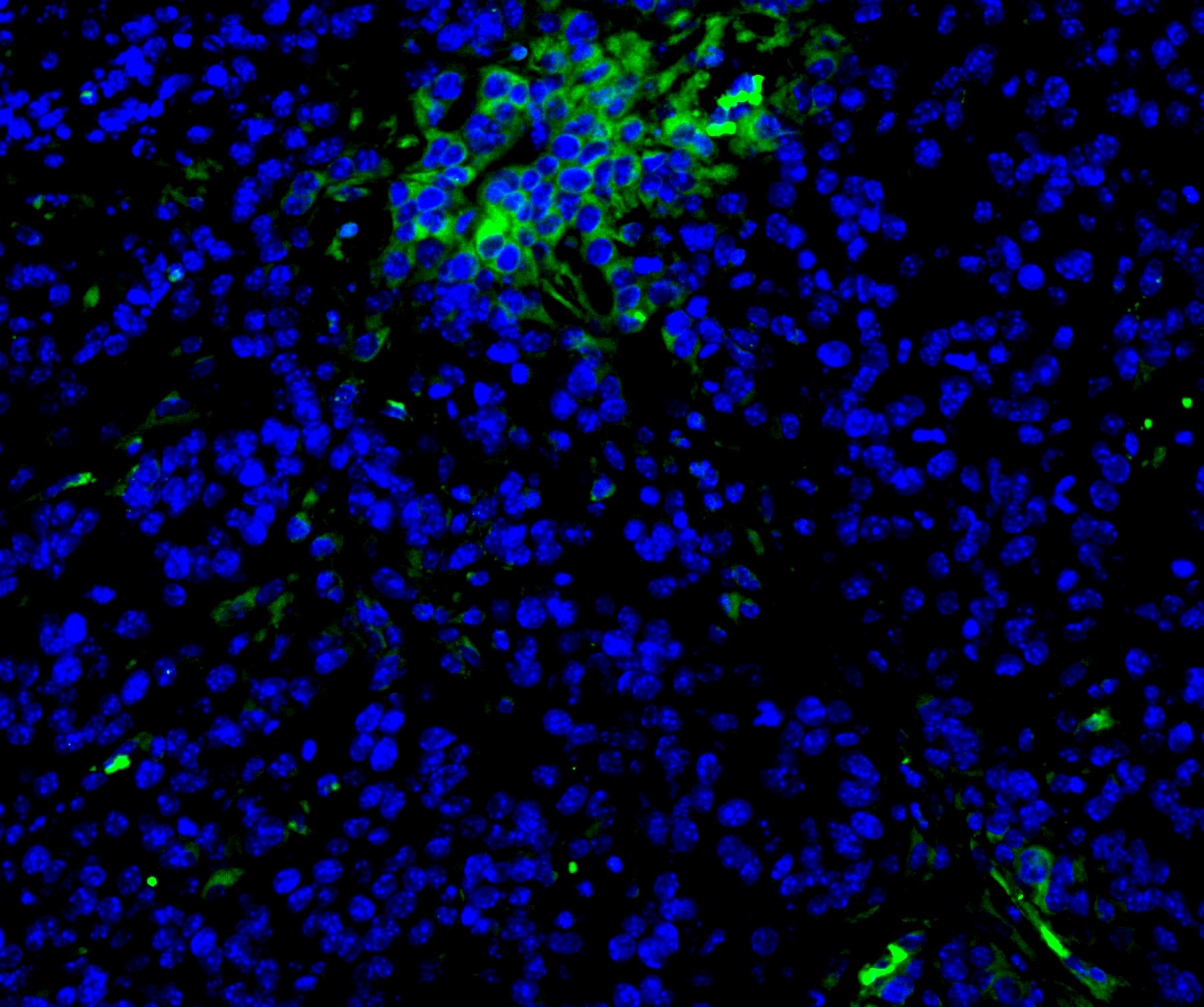
Mouse PD-L1/B7-H1 Antibody Summary
Met1-His239
Accession # Q9EP73
Applications
Please Note: Optimal dilutions should be determined by each laboratory for each application. General Protocols are available in the Technical Information section on our website.
Scientific Data
 View Larger
Detection of Mouse PD-L1/B7-H1 by Western Blot. Western blot shows lysates of RAW 264.7 mouse monocyte/macrophage cell line and J774A.1 mouse reticulum cell sarcoma macrophage cell line untreated (-) or treated (+) with 10 µg/mL LPS for 4 hours. PVDF membrane was probed with 2 µg/mL of Rabbit Anti-Mouse PD-L1/B7-H1 Monoclonal Antibody (Catalog # MAB90781) followed by HRP-conjugated Anti-Rabbit IgG Secondary Antibody (Catalog # HAF008). A specific band was detected for PD-L1/B7-H1 at approximately 50-55 kDa (as indicated). This experiment was conducted under reducing conditions and using Immunoblot Buffer Group 1.
 View Larger
Detection of PD-L1/B7-H1 in RAW 264.7 Mouse Cell Line by Flow Cytometry. RAW 264.7 mouse monocyte/macrophage cell line either treated with LPS overnight (filled histogram) or untreated (open histogram) was stained with Rabbit Anti-Mouse PD-L1/B7-H1 Monoclonal Antibody (Catalog # MAB90781), followed by Allophycocyanin-conjugated Anti-Rabbit IgG Secondary Antibody (Catalog # F0111). View our protocol for Staining Membrane-associated Proteins.
 View Larger
Detection of PD-L1/B7-H1 in HEK293 Human Cell Line Transfected with Mouse PD-L1/B7-H1 and eGFP by Flow Cytometry. HEK293 human embryonic kidney cell line transfected with either (A) mouse PD-L1/B7-H1 or (B) irrelevant transfectants and eGFP was stained with Rabbit Anti-Mouse PD-L1/B7-H1 Monoclonal Antibody (Catalog # MAB90781) followed by Allophycocyanin-conjugated Anti-Rabbit IgG Secondary Antibody (Catalog # F0111). Quadrant markers were set based on control antibody staining (Catalog # MAB1050). View our protocol for Staining Membrane-associated Proteins.
 View Larger
PD-L1/B7-H1 in Mouse Thymus. PD-L1/B7-H1 was detected in perfusion fixed frozen sections of mouse thymus using Rabbit Anti-Mouse PD-L1/B7-H1 Monoclonal Antibody (Catalog # MAB90781) at 5 µg/mL for 1 hour at room temperature followed by incubation with the Anti-Rabbit IgG VisUCyte™ HRP Polymer Antibody (Catalog # VC003). Tissue was stained using DAB (brown) and counterstained with hematoxylin (blue). Specific staining was localized to thymocytes. View our protocol for IHC Staining with VisUCyte HRP Polymer Detection Reagents.
Reconstitution Calculator
Preparation and Storage
- 12 months from date of receipt, -20 to -70 °C as supplied.
- 1 month, 2 to 8 °C under sterile conditions after reconstitution.
- 6 months, -20 to -70 °C under sterile conditions after reconstitution.
Background: PD-L1/B7-H1
Mouse B7 homolog 1(B7-H1), also called programmed death ligand 1 (PD-L1) and programmed cell death 1 ligand 1 (PDCD1L1), is a member of the B7 family of proteins that provide signals for regulating T-cell activation and tolerance (1-4). Other family members include B7-1, B7-2, B7-H2, B7-H3 and PD-L2. B7 proteins are immunoglobulin (Ig) superfamily members with extracellular Ig-V-like and Ig-C-like domains and a short cytoplasmic region. Among the family members, they share from 20-40% amino acid (aa) sequence identity. The cloned mouse B7-H1/PD-L1 cDNA encodes a 290 aa type I membrane precursor protein with a putative 18 aa signal peptide, a 220 aa extracellular region containing one V-like and one C-like Ig domain, a 22 aa transmembrane region, and a 30 aa cytoplasmic domain. Mouse and human B7-H1/PD-L1 share approximately 70% aa sequence identity. B7-H1/PD-L1 is one of two ligands for programmed death-1 (PD-1), a member of the CD28 family of immunoreceptors. The other identified ligand is PD-L2. Mouse B7-H1/PD-L1 and PD-L2 share approximately 34% aa sequence identity and have similar functions. B7-H1/PD-L1 is constitutively expressed in various lymphoid and non-lymphoid organs including placenta, heart, pancreas, lung, liver, and endothelium
(1‑4). The expression of B7-H1/PD-L1 is detected on B cells, T cells, monocytes, dendritic cells and thymic epithelial cells. IFN-gamma treatment induces B7-H1/PD-L1 expression in monocytes, dendritic cells, and endothelial cells. B7-H1/PD-L1 expression is also upregulated in a variety of tumor cell lines. On previously activated T cells, B7-H1/PD-L1 interaction with PD-1 inhibits TCR-mediated proliferation and cytokine production, suggesting an inhibitory role in regulating immune responses. In contrast, a costimulatory function for the PD-1 ligands on resting T cells has also beenreported (1-4).
- Tamura, H. et al. (2001) Blood 97:1809.
- Freeman, G. et al. (2000) J. Exp. Med. 192:1027.
- Sharpe, A.H. and G. J. Freeman (2002) Nat. Rev. Immunol. 2:116.
- Coyle, A. and J. Gutierrez-Ramos (2001) Nat. Immunol. 2:203.
Product Datasheets
Citations for Mouse PD-L1/B7-H1 Antibody
R&D Systems personnel manually curate a database that contains references using R&D Systems products. The data collected includes not only links to publications in PubMed, but also provides information about sample types, species, and experimental conditions.
12
Citations: Showing 1 - 10
Filter your results:
Filter by:
-
Enhanced bypass of PD-L1 translation reduces the therapeutic response to mTOR kinase inhibitors
Authors: Cao, Y;Ye, Q;Ma, M;She, QB;
Cell reports
Species: Mouse
Sample Types: Cell Lysates
Applications: Western Blot -
CDK4/6 inhibitors and the pRB-E2F1 axis suppress PVR and PD-L1 expression in triple-negative breast cancer
Authors: Shrestha, M;Wang, DY;Ben-David, Y;Zacksenhaus, E;
Oncogenesis
Species: Mouse
Sample Types: Cell Lysates
Applications: Western Blot -
PD-L1 induction via the MEK-JNK-AP1 axis by a neddylation inhibitor promotes cancer-associated immunosuppression
Authors: S Zhang, X You, T Xu, Q Chen, H Li, L Dou, Y Sun, X Xiong, MA Meredith, Y Sun
Cell Death & Disease, 2022-10-03;13(10):844.
Species: Mouse
Sample Types: In Vivo
Applications: Neutralization -
Engineered immunomodulatory accessory cells improve experimental allogeneic islet transplantation without immunosuppression
Authors: X Wang, K Wang, M Yu, D Velluto, X Hong, B Wang, A Chiu, JM Melero-Mar, AA Tomei, M Ma
Science Advances, 2022-07-22;8(29):eabn0071.
Species: Mouse
Sample Types: Whole Cells
Applications: ICC -
Colorectal Cancer Progression Is Potently Reduced by a Glucose-Free, High-Protein Diet: Comparison to Anti-EGFR Therapy
Authors: K Skibbe, AK Brethack, A Sünderhauf, M Ragab, A Raschdorf, M Hicken, H Schlichtin, J Preira, J Brandt, D Castven, B Föh, R Pagel, JU Marquardt, C Sina, S Derer
Cancers, 2021-11-19;13(22):.
Species: Mouse
Sample Types: Cell Lysates
Applications: Western Blot -
The role of PD-1/PD-L1 checkpoint in arsenic lung tumorigenesis
Authors: W Xu, J Cui, L Wu, C He, G Chen
Toxicology and Applied Pharmacology, 2021-06-22;426(0):115633.
Species: Mouse
Sample Types: Whole Tissue
Applications: IHC -
Knockdown of CDK5 down-regulates PD-L1 via the ubiquitination-proteasome pathway and improves antitumor immunity in lung adenocarcinoma
Authors: L Gao, L Xia, W Ji, Y Zhang, W Xia, S Lu
Translational Oncology, 2021-06-12;14(9):101148.
Species: Mouse
Sample Types: Cell Lysates
Applications: Western Blot -
Human Biofield Therapy and the Growth of Mouse Lung Carcinoma
Authors: P Yang, Y Jiang, PR Rhea, T Coway, D Chen, M Gagea, SL Harribance, L Cohen
Integr Cancer Ther, 2019-01-01;18(0):1534735419840.
Species: Mouse
Sample Types: Tissue Homogenates
Applications: Western Blot -
The Circadian Clock Controls Immune Checkpoint Pathway in Sepsis
Authors: W Deng, S Zhu, L Zeng, J Liu, R Kang, M Yang, L Cao, H Wang, TR Billiar, J Jiang, M Xie, D Tang
Cell Rep, 2018-07-10;24(2):366-378.
Species: Mouse
Sample Types: Cell Lysates
Applications: Western Blot -
Inhibition of LSD1 with Bomedemstat Sensitizes Small Cell Lung Cancer to Immune Checkpoint Blockade and T-Cell Killing
Authors: Joseph B. Hiatt, Holly Sandborg, Sarah M. Garrison, Henry U. Arnold, Sheng-You Liao, Justin P. Norton et al.
Clinical Cancer Research
-
Cyclin D–CDK4 kinase destabilizes PD-L1 via cullin 3–SPOP to control cancer immune surveillance
Authors: Jinfang Zhang, Xia Bu, Haizhen Wang, Yasheng Zhu, Yan Geng, Naoe Taira Nihira et al.
Nature
-
Intestinal TMEM16A control luminal chloride secretion in a NHERF1 dependent manner
Authors: Ling X, Han W, Jiang X, et al.
Biomaterials
FAQs
No product specific FAQs exist for this product, however you may
View all Antibody FAQsReviews for Mouse PD-L1/B7-H1 Antibody
Average Rating: 4.4 (Based on 8 Reviews)
Have you used Mouse PD-L1/B7-H1 Antibody?
Submit a review and receive an Amazon gift card.
$25/€18/£15/$25CAN/¥75 Yuan/¥2500 Yen for a review with an image
$10/€7/£6/$10 CAD/¥70 Yuan/¥1110 Yen for a review without an image
Filter by:
Tissue lysates from mammary gland tumors

