Mouse/Rat/Canine/Porcine TGF-beta 2 Quantikine ELISA Kit Summary
Product Summary
Recovery
The recovery of TGF-beta 2 spiked to levels throughout the range of the assay in various matrices was evaluated.
| Sample Type | Average % Recovery | Range % |
|---|---|---|
| Cell Culture Supernates (n=4) | 92 | 80-102 |
| Cell Lysates (n=2) | 94 | 86-98 |
| EDTA Plasma (n=4) | 84 | 78-94 |
| Heparin Plasma (n=4) | 82 | 74-92 |
| Serum (n=4) | 98 | 85-114 |
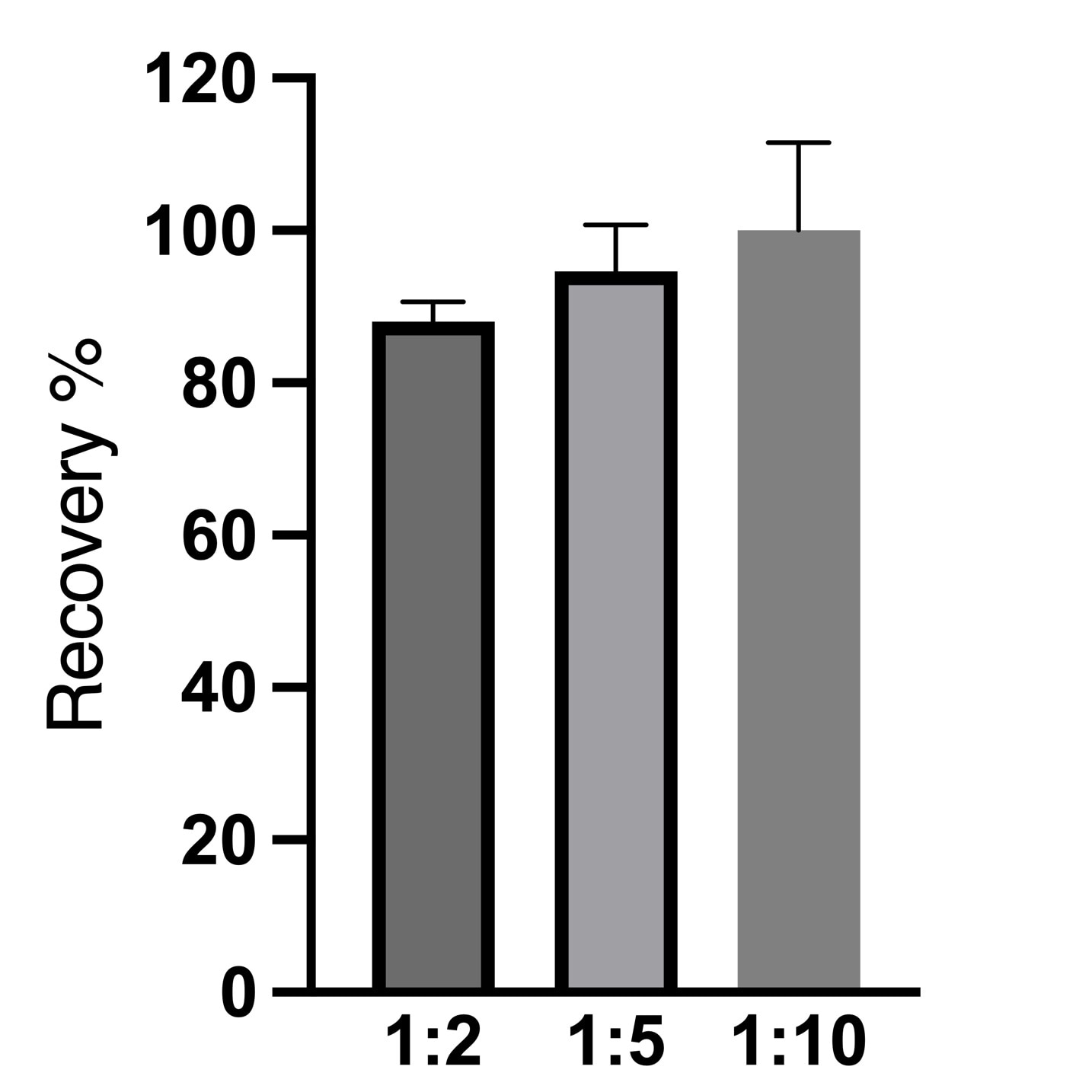
Linearity
Scientific Data
Product Datasheets
Preparation and Storage
Background: TGF-beta 2
TGF-beta 2 (Transforming Growth Factor beta 2) is one of three closely related mammalian members of the large TGF-beta superfamily that share a characteristic cysteine knot structure (1-7). TGF-beta 1, 2 and 3 are encoded by separate genes, but are often called isoforms. They are highly pleiotropic cytokines that are proposed to act as cellular switches regulating processes such as immune function, proliferation and epithelial-mesenchymal transition (4-8). Mammalian TGF-beta 2 is secreted as a 395 amino acid (aa) proprotein that is processed by a furin-like convertase to generate an N-terminal latency-associated peptide (LAP, ~232 aa) and a C-terminal mature TGF-beta 2 (~112 aa) that remain associated via hydrogen bonding (9-13). Serine proteases such as plasmin, matrix metalloproteases, or thrombospondin-1, along with cofactors such as certain integrins, can dissociate LAP and release active TGF-beta 2 (11-14). In many types of cells, latent TGF-beta binding protein (LTBP) is covalently linked to the LAP homodimer prior to secretion. LTBP creates a large latent complex that is secreted, but may bind and localize to the extracellular matrix (10, 11). For TGF-beta isoforms, the latency components act as natural antagonists of TGF-beta activity, target TGF-beta to distinct tissues, and maintain a reservoir of TGF-beta (1). Mature mouse and rat TGF-beta 2 share 100% aa sequence identity, and share 97% aa identity with human, porcine, canine, equine and bovine TGF-beta 2.
Assay Procedure
Refer to the product- Prepare all reagents, standard dilutions, and samples as directed in the product insert.
- Remove excess microplate strips from the plate frame, return them to the foil pouch containing the desiccant pack, and reseal.
- Add 50 µL of Assay Diluent to each well.
- Add 50 µL of Standard, Control, or sample to each well. Cover with a plate sealer, and incubate at room temperature for 2 hours on a horizontal orbital microplate shaker.
- Aspirate each well and wash, repeating the process 3 times for a total of 4 washes.
- Add 100 µL of Conjugate to each well. Cover with a new plate sealer, and incubate at room temperature for 2 hours on the shaker.
- Aspirate and wash 4 times.
- Add 100 µL Substrate Solution to each well. Incubate at room temperature for 30 minutes on the benchtop. PROTECT FROM LIGHT.
- Add 100 µL of Stop Solution to each well. Read at 450 nm within 30 minutes. Set wavelength correction to 540 nm or 570 nm.


Citations for Mouse/Rat/Canine/Porcine TGF-beta 2 Quantikine ELISA Kit
R&D Systems personnel manually curate a database that contains references using R&D Systems products. The data collected includes not only links to publications in PubMed, but also provides information about sample types, species, and experimental conditions.
14
Citations: Showing 1 - 10
Filter your results:
Filter by:
-
Therapeutic role of mesenchymal stem cells and platelet-rich plasma on skin burn healing and rejuvenation: A focus on scar regulation, oxido-inflammatory stress and apoptotic mechanisms
Authors: Tammam, BMH;Habotta, OA;El-Khadragy, M;Abdel Moneim, AE;Abdalla, MS;
Heliyon
Species: Rat
Sample Types: Tissue Homogenates
-
alpha1A Adrenoreceptor blockade attenuates myocardial infarction by modulating the integrin-linked kinase/TGF-beta/Smad signaling pathways
Authors: NM Alrasheed, RB Alammari, TK Alshammari, MA Alamin, AO Alharbi, AS Alonazi, AF Bin Dayel, NM Alrasheed
BMC cardiovascular disorders, 2023-03-24;23(1):153.
Species: Rat
Sample Types: Serum
-
Regenerative Effect of Mesenchymal Stem Cell on Cartilage Damage in a Porcine Model
Authors: Lin, S;Panthi, S;Hsuuw, Y;Chen, S;Huang, M;Sieber, M;Hsuuw, Y;
Biomedicines
Species: Porcine
Sample Types: Serum
-
Fibrillin-1 mutant mouse captures defining features of human primary open glaucoma including anomalous aqueous humor TGF beta-2
Authors: MK Ko, JI Woo, JM Gonzalez, G Kim, L Sakai, J Peti-Peter, JA Kelber, YK Hong, JC Tan
Oncogene, 2022-06-23;12(1):10623.
Species: Mouse
Sample Types: Aqueous Humor
-
EZH2-mediated H3K27me3 promotes autoimmune hepatitis progression by regulating macrophage polarization
Authors: G Chi, JH Pei, XQ Li
International immunopharmacology, 2022-02-19;106(0):108612.
Species: Mouse
Sample Types: Cell Culture Supernates
-
Microglia-derived interleukin-10 accelerates post-intracerebral hemorrhage hematoma clearance by regulating CD36
Authors: Q Li, X Lan, X Han, F Durham, J Wan, A Weiland, RC Koehler, J Wang
Brain, Behavior, and Immunity, 2021-02-13;94(0):437-457.
Species: Mouse
Sample Types: Tissue Homogenates
-
Role of epidermal growth factor receptor inhibitor-induced interferon pathway signaling in the head and neck squamous cell carcinoma therapeutic response
Authors: SP Korpela, TK Hinz, A Oweida, J Kim, J Calhoun, R Ferris, RA Nemenoff, SD Karam, ET Clambey, LE Heasley
Journal of Translational Medicine, 2021-01-23;19(1):43.
Species: Mouse
Sample Types: Cell Culture Supernates
-
Analysis of structural components of decellularized scaffolds in renal fibrosis
Authors: R Zhang, J Jiang, Y Yu, F Wang, N Gao, Y Zhou, X Wan, Z Wang, P Wei, J Mei
Bioactive materials, 2021-01-16;6(7):2187-2197.
Species: Rat
Sample Types: Cell Culture Supernates
-
GL261 luciferase-expressing cells elicit an anti-tumor immune response: an evaluation of murine glioma models
Authors: VE Sanchez, JP Lynes, S Walbridge, X Wang, NA Edwards, AK Nwankwo, HP Sur, GA Dominah, A Obungu, N Adamstein, PK Dagur, D Maric, J Munasinghe, JD Heiss, EK Nduom
Sci Rep, 2020-07-03;10(1):11003.
Species: Mouse
Sample Types: Cell Culture Supernates
-
De novo design of functional zwitterionic biomimetic material for immunomodulation
Authors: B Li, Z Yuan, P Jain, HC Hung, Y He, X Lin, P McMullen, S Jiang
Sci Adv, 2020-05-29;6(22):eaba0754.
Species: Mouse
Sample Types: Cell Culture Supernates
-
Evaluation of Neurosecretome from Mesenchymal Stem Cells Encapsulated in Silk Fibroin Hydrogels
Authors: Y Martín-Mar, L Fernández-, MH Sanchez-Re, N Marí-Buyé, FJ Rojo, J Pérez-Rigu, M Ramos, GV Guinea, F Panetsos, D González-N
Sci Rep, 2019-06-19;9(1):8801.
Species: Mouse
Sample Types: Cell Culture Supernates
-
TGF-?3 Promotes MUC5AC Hyper-Expression by Modulating Autophagy Pathway in Airway Epithelium
Authors: Y Zhang, H Tang, X Yuan, Q Ran, X Wang, Q Song, L Zhang, Y Qiu, X Wang
EBioMedicine, 2018-07-08;0(0):.
Species: Mouse
Sample Types: BALF
-
TGF-beta3-expressing CD4+CD25(-)LAG3+ regulatory T cells control humoral immune responses.
Authors: Okamura T, Sumitomo S, Morita K, Iwasaki Y, Inoue M, Nakachi S, Komai T, Shoda H, Miyazaki J, Fujio K, Yamamoto K
Nat Commun, 2015-02-19;6(0):6329.
Species: Mouse
Sample Types: Cell Culture Supernates
-
Resolvin D1 and lipoxin A4 improve alveolarization and normalize septal wall thickness in a neonatal murine model of hyperoxia-induced lung injury.
Authors: Martin C, Zaman M, Gilkey C, Salguero M, Hasturk H, Kantarci A, Van Dyke T, Freedman S
PLoS ONE, 2014-06-03;9(6):e98773.
Species: Mouse
Sample Types: Tissue Homogenates
FAQs
No product specific FAQs exist for this product, however you may
View all ELISA FAQsReviews for Mouse/Rat/Canine/Porcine TGF-beta 2 Quantikine ELISA Kit
Average Rating: 5 (Based on 2 Reviews)
Have you used Mouse/Rat/Canine/Porcine TGF-beta 2 Quantikine ELISA Kit?
Submit a review and receive an Amazon gift card.
$25/€18/£15/$25CAN/¥75 Yuan/¥2500 Yen for a review with an image
$10/€7/£6/$10 CAD/¥70 Yuan/¥1110 Yen for a review without an image
Filter by:


