Mouse TSG-6 Antibody Summary
Trp18-Leu275
Accession # O08859
Applications
Please Note: Optimal dilutions should be determined by each laboratory for each application. General Protocols are available in the Technical Information section on our website.
Scientific Data
 View Larger
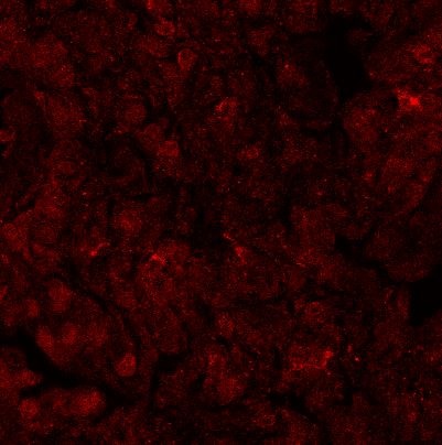
TSG‑6 in Mouse Splenocytes. TSG-6 was detected in immersion fixed mouse splenocytes using Goat Anti-Mouse TSG-6 Antigen Affinity-purified Polyclonal Antibody (Catalog # AF2326) at 15 µg/mL for 3 hours at room temperature. Cells were stained using the NorthernLights™ 557-conjugated Anti-Goat IgG Secondary Antibody (red; Catalog # NL001) and counterstained with DAPI (blue). Specific staining was localized to cytoplasm. View our protocol for Fluorescent ICC Staining of Non-adherent Cells.
Reconstitution Calculator
Preparation and Storage
- 12 months from date of receipt, -20 to -70 °C as supplied.
- 1 month, 2 to 8 °C under sterile conditions after reconstitution.
- 6 months, -20 to -70 °C under sterile conditions after reconstitution.
Background: TSG-6
TSG-6 (TNF-stimulated gene 6; also named TNFIP6) is a secreted, 35‑39 kDa group A member of the LINK-Module superfamily of proteins (1‑4). Mouse TSG-6 is synthesized as a 275 amino acid (aa) precursor. It contains a 17 aa signal sequence and a 258 aa mature region (5, 6). The mature region has an N-terminal link module (aa 36‑129) and a C-terminal CUB (C1s/C1r; urchin embryonic growth factor; BMP1) domain (aa 135‑246). Link modules bind hyaluronan (HA) and participate in extracellular matrix (ECM) assembly (7). Mature mouse TSG-6 shares 97%, 94% and 94% aa identity with rat, human and canine TSG-6, respectively. Cells reported to express TGF-6 include activated fibroblasts, synoviocytes, chondrocytes, neutrophils, proximal tubular epithelium, bronchial epithelium, endothelium, and visceral plus vascular smooth muscle (2, 8). TSG-6 has multiple functions, many of which involve the ECM. It is suggested to stabilize HA‑rich ECM. It does so by serving as an intermediary, or as a link between the individual subunits of the extracellular decameric pentraxin 3 and the surrounding hyaluronan matrix (9). It also provides structure and organization to hyaluronan. This is accomplished by a TSG-6 mediated transfer of an 80‑85 kDa protein subunit from I alpha I (inter-alpha -inhibitor) to HA. I alpha I is a four-component, 225 kDa serine protease inhibitor. It contains a protease inhibitor subunit (bikunin), two independent, accompaning protein chains (HC1 and HC2), and a short chondroitin sulfate linking moiety. TSG-6 is a catalyst for the removal and transient binding of either HC chain. Each chain is subsequently transferred and covalently-linked to the surrounding HA. This provides substance and reinforcement to the ECM (1, 2, 10, 11, 12). This disassembly of I alpha I also leads to free bikunin, which in the “free” state becomes a potent inhibitor of serine proteases (8).
- Milner, C.M. et al. (2006) Biochem. Soc. Trans. 34:446.
- Milner, C.M. and A.J. Day (2003) J. Cell Sci. 116:1863.
- Wisnieewski, H-G. and J. Vilcek (2004) Cytokine Growth Factor Rev. 15:129.
- Blundell, C.D. et al. (2005) J. Biol. Chem. 280:18189.
- Fulop, C. et al. (1997) Gene 202:95.
- Fulop, C. et al. (2003) Development 130:2253.
- Kohda, D. et al. (1996) Cell 86:767.
- Forteza, R. et al. (2007) Am. J. Respir. Cell Mol. Biol. 36:20.
- Salustri, A. et al. (2003) Development 131:1577.
- Rugg, M.S. et al. (2005) J. Biol. Chem. 280:25674.
- Sanggaard, K.W. et al. (2006) Biochemistry 45:7661.
- Sanggaard, K.W. et al. (2005) J. Biol. Chem. 280:11936.
Product Datasheets
Citations for Mouse TSG-6 Antibody
R&D Systems personnel manually curate a database that contains references using R&D Systems products. The data collected includes not only links to publications in PubMed, but also provides information about sample types, species, and experimental conditions.
3
Citations: Showing 1 - 3
Filter your results:
Filter by:
-
Mesenchymal Stromal Cells Inhibit Neutrophil Effector Functions in a Murine Model of Ocular Inflammation
Authors: SK Mittal, A Mashaghi, A Amouzegar, M Li, W Foulsham, SK Sahu, SK Chauhan
Invest. Ophthalmol. Vis. Sci., 2018-03-01;59(3):1191-1198.
Species: Mouse
Sample Types: Whole Cells
Applications: Neutralization -
Hyaluronan and Hyaluronan Binding Proteins Are Normal Components of Mouse Pancreatic Islets and Are Differentially Expressed by Islet Endocrine Cell Types
Authors: Rebecca L. Hull, Pamela Y. Johnson, Kathleen R. Braun, Anthony J. Day, Thomas N. Wight
Journal of Histochemistry & Cytochemistry
-
A Retrospective Analysis of the Cartilage Kunitz Protease Inhibitory Proteins Identifies These as Members of the Inter-?-Trypsin Inhibitor Superfamily with Potential Roles in the Protection of the Articulatory Surface
Authors: SM Smith, J Melrose
Int J Mol Sci, 2019-01-24;20(3):.
FAQs
No product specific FAQs exist for this product, however you may
View all Antibody FAQsReviews for Mouse TSG-6 Antibody
Average Rating: 3 (Based on 1 Review)
Have you used Mouse TSG-6 Antibody?
Submit a review and receive an Amazon gift card.
$25/€18/£15/$25CAN/¥75 Yuan/¥2500 Yen for a review with an image
$10/€7/£6/$10 CAD/¥70 Yuan/¥1110 Yen for a review without an image
Filter by:


