Mouse Uromodulin Antibody Summary
Ser24-Ala618
Accession # Q91X17
*Small pack size (-SP) is supplied either lyophilized or as a 0.2 µm filtered solution in PBS.
Applications
Please Note: Optimal dilutions should be determined by each laboratory for each application. General Protocols are available in the Technical Information section on our website.
Scientific Data
 View Larger
Detection of Mouse Uromodulin by Western Blot. Western blot shows lysates of mouse kidney tissue. PVDF membrane was probed with 1 µg/mL of Sheep Anti-Mouse Uromodulin Antigen Affinity-purified Polyclonal Antibody (Catalog # AF5175) followed by HRP-conjugated Anti-Sheep IgG Secondary Antibody (HAF016). A specific band was detected for Uromodulin at approximately 115 kDa (as indicated). This experiment was conducted under reducing conditions and using Immunoblot Buffer Group 8.
 View Larger
Uromodulin in Mouse Kidney. Uromodulin was detected in immersion fixed frozen sections of mouse kidney using Sheep Anti-Mouse Uromodulin Antigen Affinity-purified Polyclonal Antibody (Catalog # AF5175) at 10 µg/mL overnight at 4 °C. Tissue was stained using the NorthernLights™ 557-conjugated Anti-Sheep IgG Secondary Antibody (red; NL010) and counterstained with DAPI (blue). Specific staining was localized to convoluted tubule epithelial cells. View our protocol for Fluorescent IHC Staining of Frozen Tissue Sections.
 View Larger
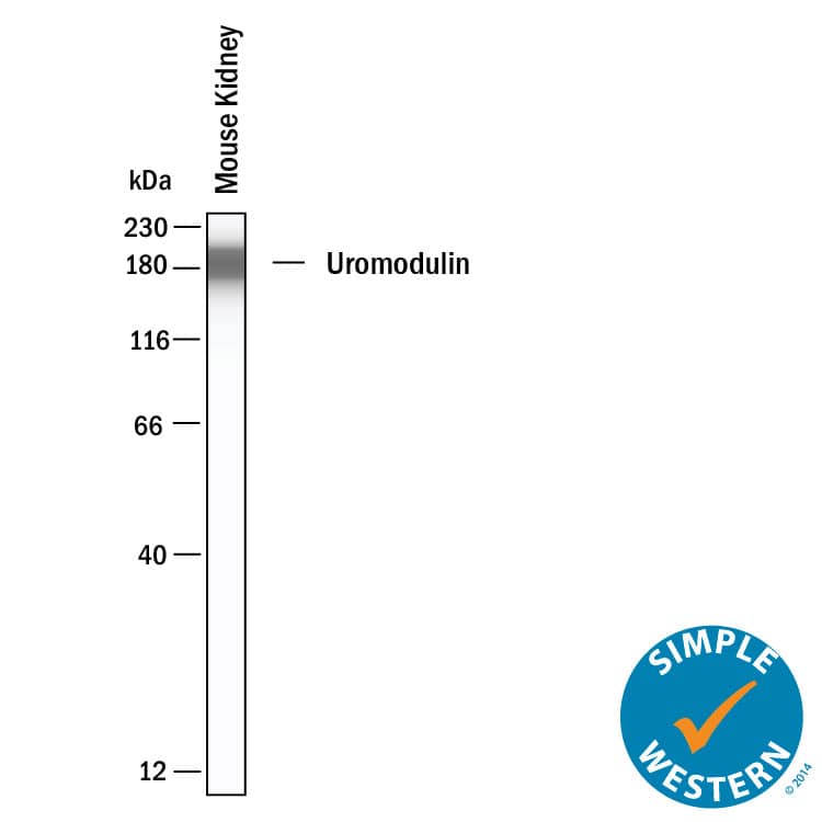
Detection of Mouse Uromodulin by Simple WesternTM. Simple Western shows lysates of mouse kidney, loaded at 0.5 mg/ml. A specific band was detected for Uromodulin at approximately 182 kDa (as indicated) using 20 µg/mL of Sheep Anti-Mouse Uromodulin Antigen Affinity-purified Polyclonal Antibody (Catalog # AF5175). This experiment was conducted under reducing conditions and using the 12-230kDa separation system.
 View Larger
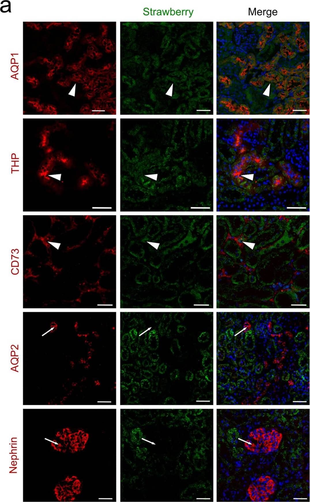
Detection of Mouse Uromodulin by Immunocytochemistry/Immunofluorescence Intraparenchymal delivery of lentiviral vectors preferentially infects epithelial cells of both kidneys and urinary bladder.(a) Confocal images of Strawberry expression in renal sections of ELS infected kidneys 60 days post injection. Sections were stained with the indicated antibodies and merged images with DAPI (blue) are presented. Preferential transduction was observed in proximal (AQP1) and distal (THP) renal tubular epithelial cells and renal fibroblasts (CD73) (white arrowheads). Strawberry was not localised to collecting ducts (AQP2) or podocytes (Nephrin) (white arrows). Scale bars, 50 μm. (b) Confocal images of Strawberry expression in the liver, spleen, lung, urinary bladder and left and right kidney of ELS infected animals 60 days post left intrarenal injection (top panels) and uninjected control animals (bottom panels). Sections were stained with an antibody against Strawberry (green) and merged images with DAPI (blue) are presented. Scale bars, 50 μm. Image collected and cropped by CiteAb from the following publication (https://pubmed.ncbi.nlm.nih.gov/26046460), licensed under a CC-BY license. Not internally tested by R&D Systems.
 View Larger
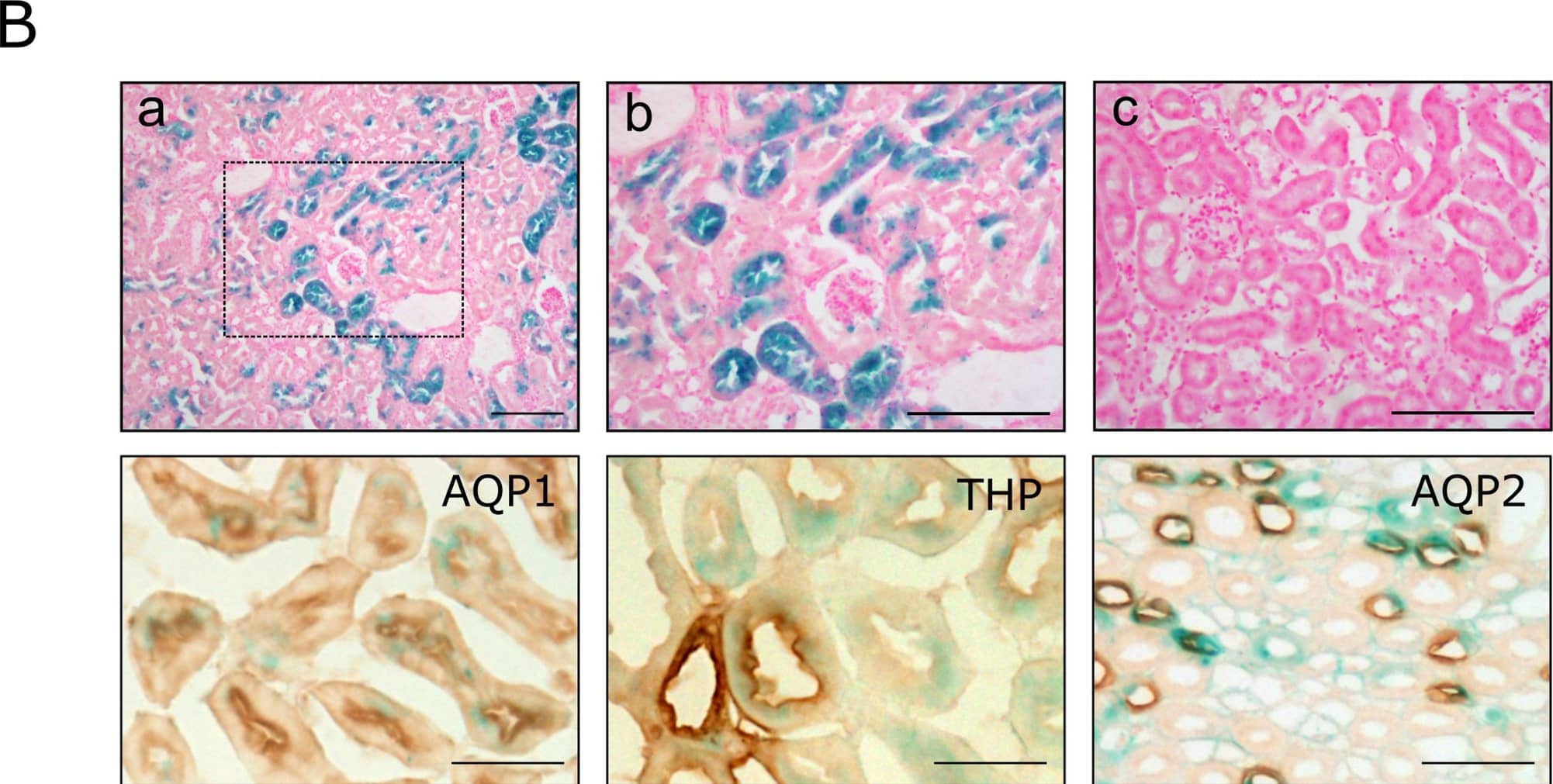
Detection of Mouse Uromodulin by Immunohistochemistry Pax8-CreERT2 mediated beta -galactosidase expression.(A) Enzymatic X-gal staining of cryosections of kidneys (top panels) derived from 10 week old Pax8-CreERT2/Rosa26R mice either induced with tamoxifen (a, renal parenchyma; b, collecting ducts) or uninduced (c). Tamoxifen administration did not induce beta -galactosidase expression in any of the other major organs examined (representative tissues presented in bottom panels). (B) beta -galactosidase positivity was exclusively found in the tubular epithelium of the kidney of tamoxifen treated mice, with no staining observed in glomeruli or bloods vessels (a, framed area is shown enlarged in b). Kidneys from vehicle treated mice were negative (c). Co-localisation studies revealed X-gal expression within tubules of all renal tubular compartment (proximal tubules—AQP1, distal tubules–THP and collecting ducts–AQP2). Scale bars, 100μm. Image collected and cropped by CiteAb from the following publication (https://pubmed.ncbi.nlm.nih.gov/26866916), licensed under a CC-BY license. Not internally tested by R&D Systems.
 View Larger
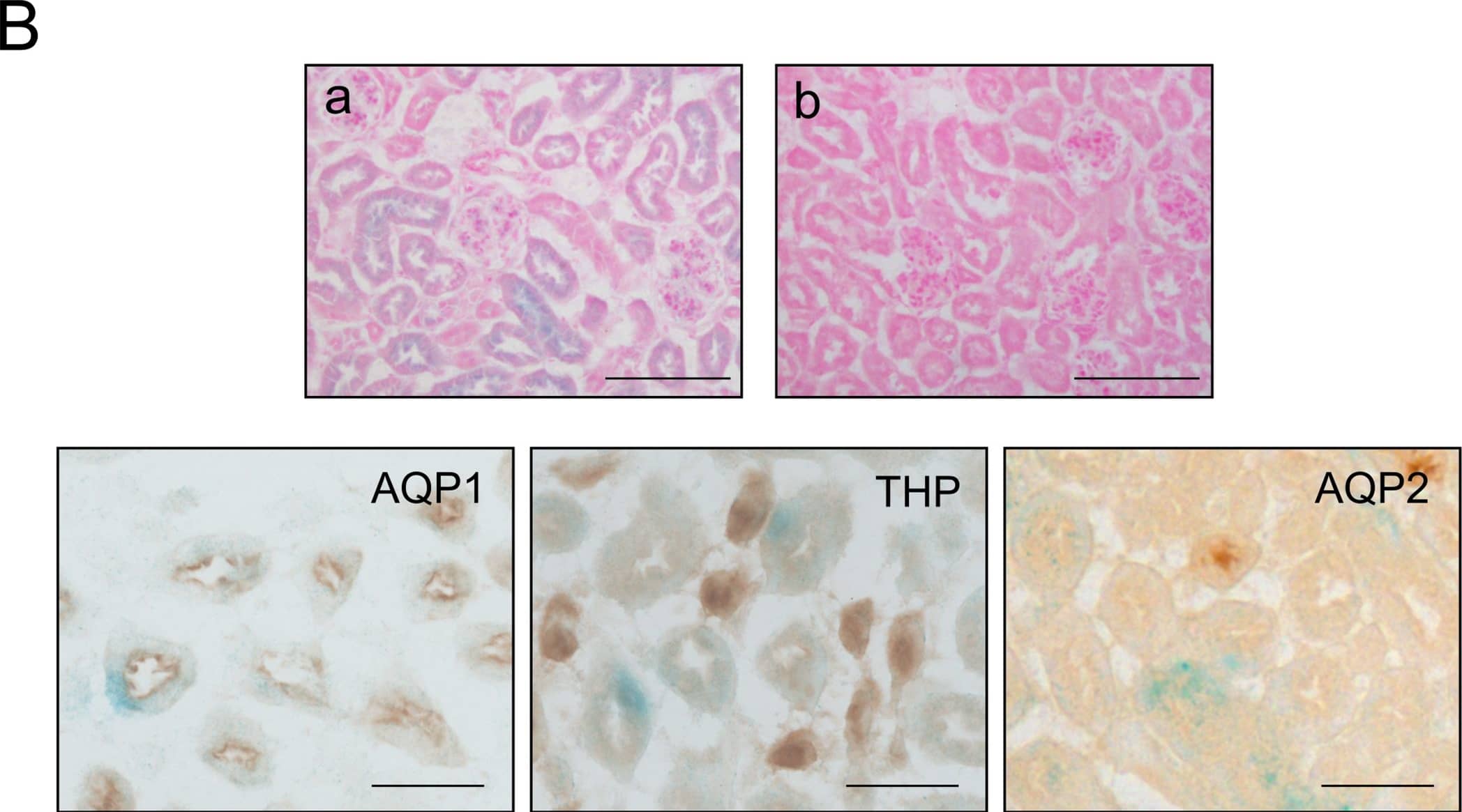
Detection of Mouse Uromodulin by Immunohistochemistry Slc22a6-CreERT2 mediated beta -galactosidase expression.(A) Enzymatic X-gal staining of cryosections of kidneys (top panels) derived from 10 week old Slc22a6-CreERT2/Rosa26R mice either induced with tamoxifen (a, renal cortex; b, medulla) or uninduced (c, renal cortex). Tamoxifen administration did not induce beta -galactosidase expression in any of the other major organs examined (representative tissues presented in bottom panels). (B) beta -galactosidase positivity was exclusively found in the tubular epithelium of the kidney of tamoxifen treated mice (a); kidneys from vehicle treated mice were negative (b). Co-localisation studies revealed specific X-gal expression within proximal tubular epithelium (AQP1) with no expression detected in distal tubules (THP) or collecting ducts (AQP2). Scale bars, 100μm. Image collected and cropped by CiteAb from the following publication (https://pubmed.ncbi.nlm.nih.gov/26866916), licensed under a CC-BY license. Not internally tested by R&D Systems.
Reconstitution Calculator
Preparation and Storage
- 12 months from date of receipt, -20 to -70 °C as supplied.
- 1 month, 2 to 8 °C under sterile conditions after reconstitution.
- 6 months, -20 to -70 °C under sterile conditions after reconstitution.
Background: Uromodulin
Uromodulin (also Tamm-Horsfall glycoprotein or THP) is a 105‑120 kDa urinary glycoprotein. It is secreted by renal tubule epithelium, acts as a binding protein for IL‑1, TNF-alpha and C1q, activates resting monocytes, and promotes neutrophil phagocytosis. Uromodulin forms high molecular weight oligomers that line the kidney tubules. Mouse Uromodulin is GPI-linked. Its proprecursor is 619 amino acids (aa) in length. It contains three EGF-like domains (aa 28‑148), a ZP domain that mediates oligomerization (aa 335‑590), and a cleavable C-terminal propeptide (aa 619‑642). There are multiple splice variants. One shows an alternate start site at Met343, and there are two substitutions, a 17 aa substitution for aa 601‑642, and a 166 aa substitution for aa 441‑642. Over aa 25‑618, mouse Uromodulin shares 78% and 89% aa identical to human and rat Uromodulin, respectively.
Product Datasheets
Citations for Mouse Uromodulin Antibody
R&D Systems personnel manually curate a database that contains references using R&D Systems products. The data collected includes not only links to publications in PubMed, but also provides information about sample types, species, and experimental conditions.
8
Citations: Showing 1 - 8
Filter your results:
Filter by:
-
Inactivation of Invs/Nphp2 in renal epithelial cells drives infantile nephronophthisis like phenotypes in mouse
Authors: Yuanyuan Li, Wenyan Xu, Svetlana Makova, Martina Brueckner, Zhaoxia Sun
eLife
-
Conserved and Divergent Molecular and Anatomic Features of Human and Mouse Nephron Patterning
Authors: Nils O. Lindström, Tracy Tran, Jinjin Guo, Elisabeth Rutledge, Riana K. Parvez, Matthew E. Thornton et al.
Journal of the American Society of Nephrology
-
LC3 lipidation is essential for TFEB activation during the lysosomal damage response to kidney injury
Authors: S Nakamura, S Shigeyama, S Minami, T Shima, S Akayama, T Matsuda, A Esposito, G Napolitano, A Kuma, T Namba-Hama, J Nakamura, K Yamamoto, M Sasai, A Tokumura, M Miyamoto, Y Oe, T Fujita, S Terawaki, A Takahashi, M Hamasaki, M Yamamoto, Y Okada, M Komatsu, T Nagai, Y Takabatake, H Xu, Y Isaka, A Ballabio, T Yoshimori
Nat Cell Biol, 2020-09-28;22(10):1252-1263.
Species: Mouse
Sample Types: Whole Cells, Whole Tissue
Applications: ICC, IHC -
Uromodulin deficiency alters tubular injury and interstitial inflammation but not fibrosis in experimental obstructive nephropathy
Authors: O Maydan, PG McDade, Y Liu, XR Wu, DG Matsell, AA Eddy
Physiol Rep, 2018-03-01;6(6):e13654.
Species: Mouse
Sample Types: Tissue Homogenates, Whole Tissue
Applications: IHC, Western Blot -
Polymerization-Incompetent Uromodulin in the Pregnant Stroke-Prone Spontaneously Hypertensive Rat
Authors: S Mary, HY Small, J Siwy, W Mullen, A Giri, C Delles
Hypertension, 2017-03-27;69(5):910-918.
Species: Rat
Sample Types: Urine
Applications: Western Blot -
Generation and Characterisation of a Pax8-CreERT2 Transgenic Line and a Slc22a6-CreERT2 Knock-In Line for Inducible and Specific Genetic Manipulation of Renal Tubular Epithelial Cells.
Authors: Espana-Agusti J, Zou X, Wong K et al.
PLoS One
-
Deletion of ADP Ribosylation Factor-Like GTPase 13B Leads to Kidney Cysts
Authors: Yuanyuan Li, Xin Tian, Ming Ma, Stephanie Jerman, Shanshan Kong, Stefan Somlo et al.
Journal of the American Society of Nephrology
-
A minimally invasive, lentiviral based method for the rapid and sustained genetic manipulation of renal tubules.
Authors: Espana-Agusti J, Tuveson DA, Adams DJ, Matakidou A.
Sci Rep
FAQs
No product specific FAQs exist for this product, however you may
View all Antibody FAQsReviews for Mouse Uromodulin Antibody
There are currently no reviews for this product. Be the first to review Mouse Uromodulin Antibody and earn rewards!
Have you used Mouse Uromodulin Antibody?
Submit a review and receive an Amazon gift card.
$25/€18/£15/$25CAN/¥75 Yuan/¥2500 Yen for a review with an image
$10/€7/£6/$10 CAD/¥70 Yuan/¥1110 Yen for a review without an image

