Rat LYVE-1 Antibody Summary
Thr53-Thr259
Accession # NP_001099756
Applications
Please Note: Optimal dilutions should be determined by each laboratory for each application. General Protocols are available in the Technical Information section on our website.
Scientific Data
 View Larger
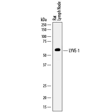
Detection of Rat LYVE‑1 by Western Blot. Western blot shows lysates of rat lymph node tissue. PVDF membrane was probed with 1 µg/mL of Sheep Anti-Rat LYVE-1 Antigen Affinity-purified Polyclonal Antibody (Catalog # AF7939) followed by HRP-conjugated Anti-Sheep IgG Secondary Antibody (Catalog # HAF016). A specific band was detected for LYVE-1 at approximately 60 kDa (as indicated). This experiment was conducted under reducing conditions and using Immunoblot Buffer Group 1.
 View Larger
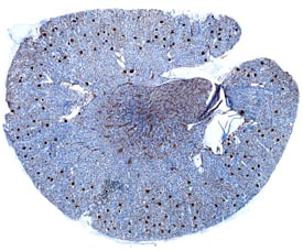
LYVE‑1 in Rat Kidney. LYVE-1 was detected in perfusion fixed frozen sections of rat kidney using Sheep Anti-Rat LYVE-1 Antigen Affinity-purified Polyclonal Antibody (Catalog # AF7939) at 1.7 µg/mL overnight at 4 °C. Tissue was stained using the Anti-Sheep HRP-DAB Cell & Tissue Staining Kit (brown; Catalog # CTS019) and counterstained with hematoxylin (blue). Specific staining was localized to glomeruli. View our protocol for Chromogenic IHC Staining of Frozen Tissue Sections.
Preparation and Storage
- 12 months from date of receipt, -20 to -70 °C as supplied.
- 1 month, 2 to 8 °C under sterile conditions after reconstitution.
- 6 months, -20 to -70 °C under sterile conditions after reconstitution.
Background: LYVE-1
LYVE-1 (Lymphatic vessel endothelial hyaluronic acid receptor 1; also CRSBP-1) is a 58-64 kDa, monomeric, glycoprotein member of the Link protein superfamily of hyaluron-binding molecules. It has limited expression, being found on the cell surface of lymphatic endothelial cells, endothelial cells of lymphoid sinuses, nodal stromal cells, and macrophages plus dendritic cells. HA (hyaluronan) is a nonsulfated, freestanding, repeating disaccharide consisting of GlcA (glucuronic acid) in beta ‑linkage with GlcNAc (N-acetylglucosamine). It should not be confused with heparan, which is sulfated, protein-linked, and composed of repeating GlcA/IdoA and GlcNAc residues in both alpha - and beta -linkages. HA is ubiquitous and occupies space between collagen fibers. It undergoes both normal, and pathology-induced turnover, and presumably does so by binding to LYVE-1, CD44 and HARE on lymphatic endothelium. Ultimately, HA is transported to the liver and nodes where it undergoes degradation. This may be necessary as low MW HA is proinflammatory. LYVE-1 is also a receptor for PDGF-BB and VEGF-A, and LYVE-1 ligation apparently induces endothelial cell contraction with the opening of intercellular junctions. Based on mouse, rat LYVE-1 is synthesized as a 343 amino acid (aa) type I transmembrane protein that contains a very long signal sequence (aa 1-52). The extracellular region is likely to be 182 aa in length (aa 53-234) and contain one Link domain (aa 62‑210). LYVE-12 is likely maintained in a default "off mode" by undergoing sialylation, possibly at Thr83. Over aa 53-259, rat LYVE-1 shares 82% and 60% aa sequence identity with mouse and human LYVE-1, respectively.
Product Datasheets
FAQs
No product specific FAQs exist for this product, however you may
View all Antibody FAQsReviews for Rat LYVE-1 Antibody
There are currently no reviews for this product. Be the first to review Rat LYVE-1 Antibody and earn rewards!
Have you used Rat LYVE-1 Antibody?
Submit a review and receive an Amazon gift card.
$25/€18/£15/$25CAN/¥75 Yuan/¥2500 Yen for a review with an image
$10/€7/£6/$10 CAD/¥70 Yuan/¥1110 Yen for a review without an image
