V5 Epitope Tag Antibody Summary
Accession # Y138513
Applications
Please Note: Optimal dilutions should be determined by each laboratory for each application. General Protocols are available in the Technical Information section on our website.
Scientific Data
 View Larger
Detection of V5 Epitope-tagged proteins by Western Blot. Western blot shows lysates of HEK293 human embryonic kidney cell line transfected with N-terminal V5-tagged CBL, N-terminal V5-tagged FSNC4, C-terminal V5-tagged LAMP2B, and C-terminal V5-tagged GALNT2. PVDF membrane was probed with 0.1 µg/mL of Rabbit Anti-V5 Epitope Tag Monoclonal Antibody (Catalog # MAB8926) followed by HRP-conjugated Anti-Rabbit IgG Secondary Antibody (Catalog # HAF008). Specific bands were detected for V5 Epitope-tagged at approximately 110 kDa, 34 kDa, 75 kDa, and 65 kDa (as indicated). This experiment was conducted under reducing conditions and using Immunoblot Buffer Group 1.
 View Larger
Detection of V5 Epitope Tag in HEK293 Human Cell Line Transfected with V5-tagged Proteins by Flow Cytometry. HEK293 human embryonic kidney cell line transfected with V5-tagged proteins was stained with Rabbit Anti-V5 Epitope Tag Monoclonal Antibody (Catalog # MAB8926, filled histogram) or isotype control antibody (Catalog # AB-105-C, open histogram), followed by Allophycocyanin-conjugated Anti-Rabbit IgG Secondary Antibody (Catalog # F0111).
 View Larger
Detection of V5 Epitope Tag by Simple WesternTM. Simple Western lane view shows lysates of HEK293 human embryonic kidney cell line transfected with N-terminal V5-tagged CBL and C-terminal V5-tagged GALNT2, loaded at 0.2 mg/mL. Specific bands were detected for V5 Epitope-tagged proteins at approximately 119 kDa and 65 kDa (as indicated) using 1 µg/mL of Rabbit Anti-V5 Epitope Tag Monoclonal Antibody (Catalog # MAB8926). This experiment was conducted under reducing conditions and using the 12-230 kDa separation system.
 View Larger
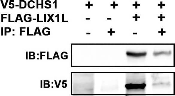
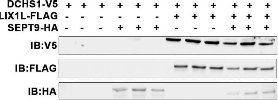
Detection of Mouse V5 Epitope Tag Antibody by Immunoprecipitation Identification of DCHS1-LIX1L-SEPT9 (DLS) protein complex. (A) Yeast two-hybrid (Y2H) screen using cytoplasmic tail (A.A. 2962–3191) of Dachsous Cadherin Related-1 (DCHS1) as bait reveals Lix1-Like (LIX1L). (B) Y2H with full-length LIX1-Like (LIX1L) identifies DCHS1 and septin-9 (SEPT9) as binding partners. In A and B, confidence scores represent likelihood of an interaction, with A being the highest level of confidence (See Methods for details). (C) Diagram depicting interacting proteins and binding domains. (D) Co-Immunoprecipitation (co-IP) and immunoblotting (IB) analysis with DCHS1-V5 and LIX1L-FLAG transfected in HEK293T cells confirms protein interaction. (E) Co-IP and IB of cVIC protein lysate treated with 10 μg peptide mimicking the LIX1L-SEPT9 binding domain (S9-FWD:5-FAM-YGRKKRRQRRR-Ahx TKWGTIEVENTTHCEFAYLRDLLIRTHMQNIKDIT-Lys(Biotin), S9-REV:5-FAM-YGRKKRRQRRR-Ahx-TIDKINQMHTRILLDRLYAFECHTTNEVEITGWKT-Lys(Biotin)). (F) IB of DCHS1-V5, LIX1L-FLAG and SEPT9-HA co-transfected into HEK293T cells depicts stabilization of DCHS1 protein only in the presence of LIX1L. Image collected and cropped by CiteAb from the following publication (https://pubmed.ncbi.nlm.nih.gov/35200715), licensed under a CC-BY license. Not internally tested by R&D Systems.
Reconstitution Calculator
Preparation and Storage
- 12 months from date of receipt, -20 to -70 °C as supplied.
- 1 month, 2 to 8 °C under sterile conditions after reconstitution.
- 6 months, -20 to -70 °C under sterile conditions after reconstitution.
Background: V5 Epitope Tag
Product Datasheets
Citations for V5 Epitope Tag Antibody
R&D Systems personnel manually curate a database that contains references using R&D Systems products. The data collected includes not only links to publications in PubMed, but also provides information about sample types, species, and experimental conditions.
5
Citations: Showing 1 - 5
Filter your results:
Filter by:
-
The ARID1A-METTL3-m6A axis ensures effective RNase H1-mediated resolution of R-loops and genome stability
Authors: Zhang, J;Chen, F;Tang, M;Xu, W;Tian, Y;Liu, Z;Shu, Y;Yang, H;Zhu, Q;Lu, X;Peng, B;Liu, X;Xu, X;Gullerova, M;Zhu, WG;
Cell reports
Species: Human
Sample Types: Cell Lysates
Applications: Immunoprecipitation -
DCHS1, Lix1L, and the Septin Cytoskeleton: Molecular and Developmental Etiology of Mitral Valve Prolapse
Authors: KS Moore, R Moore, DB Fulmer, L Guo, C Gensemer, R Stairley, J Glover, TC Beck, JE Morningsta, R Biggs, R Muhkerjee, A Awgulewits, RA Norris
Journal of cardiovascular development and disease, 2022-02-17;9(2):.
Species: Human
Sample Types: Cell Lysates
Applications: Immunoprecipitation -
Host and viral determinants for efficient SARS-CoV-2 infection of the human lung
Authors: H Chu, B Hu, X Huang, Y Chai, D Zhou, Y Wang, H Shuai, D Yang, Y Hou, X Zhang, TT Yuen, JP Cai, AJ Zhang, J Zhou, S Yuan, KK To, IH Chan, KY Sit, DC Foo, IY Wong, AT Ng, TT Cheung, SY Law, WK Au, MA Brindley, Z Chen, KH Kok, JF Chan, KY Yuen
Nature Communications, 2021-01-08;12(1):134.
Species: Hamster - Mesocricetus auratus (Golden Hamster)
Sample Types: Cell Lysates
Applications: Western Blot -
Forward genetics identifies a novel sleep mutant with sleep state inertia and REM sleep deficits
Authors: GT Banks, MCC Guillaumin, I Heise, P Lau, M Yin, N Bourbia, C Aguilar, MR Bowl, C Esapa, LA Brown, S Hasan, E Tagliatti, E Nicholson, RS Bains, S Wells, VV Vyazovskiy, K Volynski, SN Peirson, PM Nolan
Sci Adv, 2020-08-12;6(33):eabb3567.
Species: Human
Sample Types: Cell Lysates
Applications: Immunoprecipitation, Western Blot -
The Zika Virus Capsid Disrupts Corticogenesis by Suppressing Dicer Activity and miRNA Biogenesis
Authors: J Zeng, S Dong, Z Luo, X Xie, B Fu, P Li, C Liu, X Yang, Y Chen, X Wang, Z Liu, J Wu, Y Yan, F Wang, JF Chen, J Zhang, G Long, SA Goldman, S Li, Z Zhao, Q Liang
Cell Stem Cell, 2020-08-06;0(0):.
Species: Human, Mouse
Sample Types: Cell Lysates
Applications: Western Blot
FAQs
No product specific FAQs exist for this product, however you may
View all Antibody FAQsReviews for V5 Epitope Tag Antibody
There are currently no reviews for this product. Be the first to review V5 Epitope Tag Antibody and earn rewards!
Have you used V5 Epitope Tag Antibody?
Submit a review and receive an Amazon gift card.
$25/€18/£15/$25CAN/¥75 Yuan/¥2500 Yen for a review with an image
$10/€7/£6/$10 CAD/¥70 Yuan/¥1110 Yen for a review without an image

